
GTX100118 WB Image
Whole cell extract (30 ug) was separated by 10% SDS-PAGE, and the membrane was blotted with GAPDH antibody (GTX100118) diluted at 1:5000.
GTX100118 WB Image
GAPDH antibody detects GAPDH protein by western blot analysis.
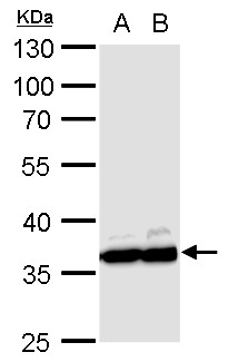
A. 30 ug PC-12 whole cell lysate/extract
B. 30 ug Rat2 whole cell lysate/extract
10% SDS-PAGE
GAPDH antibody (GTX100118) dilution: 1:10000
The HRP-conjugated anti-rabbit IgG antibody (GTX213110-01) was used to detect the primary antibody.
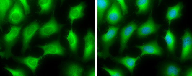
GTX100118 ICC/IF Image
GAPDH antibody detects GAPDH protein at cytoplasm by immunofluorescent analysis.Sample: HeLa cells were fixed in 4% paraformaldehyde at RT for 15 min.Green: GAPDH stained by GAPDH antibody (GTX100118) diluted at 1:500.Blue: Hoechst 33342 staining.
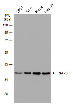
GTX100118 WB Image
Various whole cell extracts (30 ug) were separated by 10% SDS-PAGE, and the membrane was blotted with GAPDH antibody (GTX100118) diluted at 1:100000. The HRP-conjugated anti-rabbit IgG antibody (GTX213110-01) was used to detect the primary antibody.
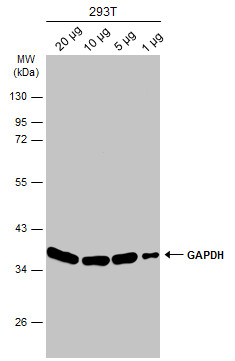
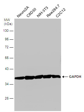
GTX100118 WB Image
Various whole cell extracts were separated by 10% SDS-PAGE, and the membrane was blotted with GAPDH antibody (GTX100118) diluted at 1:10000. The HRP-conjugated anti-rabbit IgG antibody (GTX213110-01) was used to detect the primary antibody.
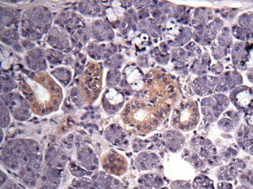
GTX100118 IHC-P Image
Immunohistochemical analysis of human salivary gland cancer, using GAPDH(GTX100118) antibody at 1:500 dilution.
GTX100118 WB Image
Various whole cell extracts (30 ug) were separated by 10% SDS-PAGE, and the membrane was blotted with GAPDH antibody (GTX100118) diluted at 1:6000. The HRP-conjugated anti-rabbit IgG antibody (GTX213110-01) was used to detect the primary antibody.
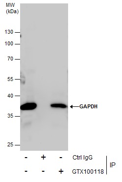
GTX100118 IP Image
Immunoprecipitation of GAPDH protein from 293T whole cell extracts using 5 ug of GAPDH antibody (GTX100118).
Western blot analysis was performed using GAPDH antibody (GTX100118).
EasyBlot anti-Rabbit IgG (GTX221666-01) was used as a secondary reagent.
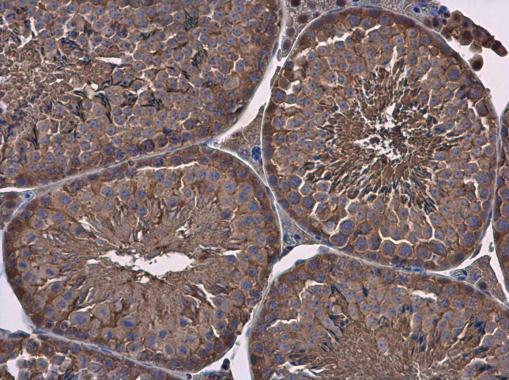
GTX100118 IHC-P Image
GAPDH antibody detects GAPDH protein at cytoplasm in mouse testis by immunohistochemical analysis.
Sample: Paraffin-embedded mouse testis.
GAPDH antibody (GTX100118) diluted at 1:500.
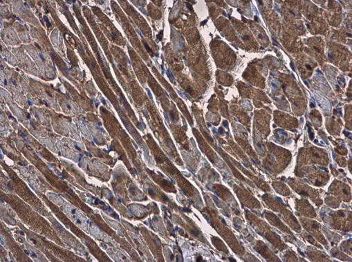
GTX100118 IHC-P Image
GAPDH antibody detects GAPDH protein at cytoplasm in rat heart by immunohistochemical analysis.
Sample: Paraffin-embedded rat heart.
GAPDH antibody (GTX100118) diluted at 1:500.