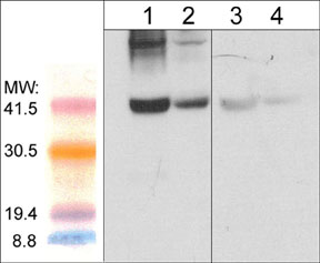
Western blot image of mouse gastrocnemius (lanes 1 & 3) and mouse diagphram tissue lysate (lanes 2 & 4). The blot was probed with anti-Atrogin-1 (AP2041; lanes 1-4) in the presence (lanes 3 & 4) or absence (lanes 1 & 2) of Atrogin-1 peptide (AX2045).
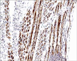
Formalin fixed, citric acid treated parafin sections of E16 mouse skeletal muscle. Sections were probed with anti-Atrogin-1 (AP2041) then anti-Rabbit:HRP before detection using DAB. (Images provided by Carl Hobbs and Dr. Pat Doherty at Wolfson Centre for Age-Related Diseases, King's College London).