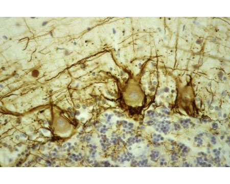
Human cerebellar cortex fixed in formalin, embedded in paraffin and stained with Mouse monoclonal antibody to Neurofilament Heavy, phosphorylated [NAP4] M-1387-50 using the ABC (avidin biotin conjugate) method. The section was counterstained with heamatoxylin-eosin (blue). This anibody stains prominent basket cell axons surrounding the large Purkinje neurons. Granule cell layer at bottom of image, molecular layer at top.