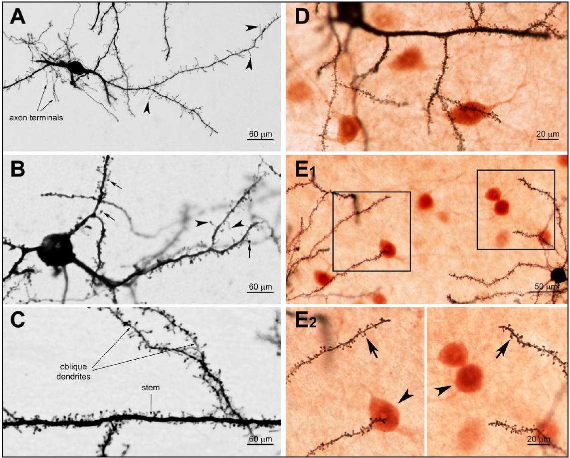
BIE(Bioenno Tech LLC/バイオエノテック)社 sliceGolgi Kit(#003760)を用いてゴルジ染色、更に免疫染色との二重染色(D~E)を行った。
A~C: 生後12日(P12)のC57BLマウスからフリーフローティング切片(厚さ100~200 um)を作製しニューロンを染色した。
(A)視床下部外側野(LHA)のニューロン。多数の樹状突起フィロポディア(矢じり部)が観察される。
(B)前頭皮質(FC)のニューロン。成熟したスパイン(矢印)や樹状突起フィロポディア(矢じり部)が見られる。
(C)前頭頭頂皮質(運動野)の錐体細胞。
D~E: ゴルジ染色と免疫染色の二重染色例。
厚さ50~100 umフリーフローティング切片をまずゴルジ染色(22±1℃で2~4日)し、その後免疫細胞染色を行った。
(D)マウス海馬台(4ヶ月齢のC57マウス)
(E1)マウス前頭頭頂皮質の体性感覚野(2ヶ月齢のC57BLマウス)
(E2)上記E1の拡大図。樹状突起スパイン(矢印)および免疫染色で標識されたニューロン(矢じり部)が見られる。
The sliceGolgi Kit was used to perform Golgi-staining alone and combined Golgi-staining and immunohistochemistry/immunocytochemistry (ICC).
A-C: The sliceGolgi Kit was used to impregnate and stain neurons in 100 ? 200 micron thick, free-floating sections of postnatal day 12 (P12) C57BL mouse.
(A) a stained neuron in the lateral hypothalamic area with numerous filopodia-like protrusions (arrowheads);
(B) a neuron in the frontal cortex with mature spines (arrows) and filopodia (arrowheads). This neuron has a giant cell body;
(C) the main (stem) and oblique dendrites of a pyramidal neuron in the motor area of the frontoparietal cortex.
D-E: The combination of Golgi-staining and ICC allows simultaneous visualization of dendritic spines and immunoreactive products.
Golgi-staining was first performed on 50?100 micron thick, free-floating sections, followed by ICC. The impregnation time was 2?4 days at 22 ± 1oC.
Images were taken from the subiculum of a 4-month old C57 mouse (D) and from the frontoparietal cortex (somatosensory area) of a 2-month old C57BL mouse (E). Boxed areas in E1 were magnified (63×) (E2) to highlight the dendritic spines (arrows) and immuno-labeled neurons (arrowheads).