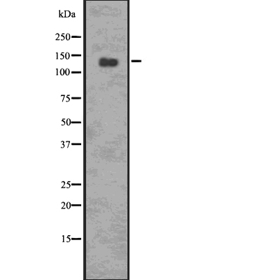
Western blot analysis of p-PERK (Thr 982) using 293 whole cell lysates
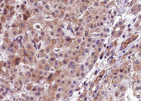
DF7576 at 1/100 staining human liver carcinoma tissue sections by IHC-P. The tissue was formaldehyde fixed and a heat mediated antigen retrieval step in citrate buffer was performed. The tissue was then blocked and incubated with the antibody for 1.5 hours at 22!aC. An HRP conjugated goat anti-rabbit antibody was used as the secondary
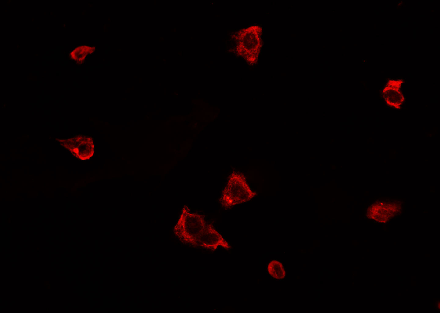
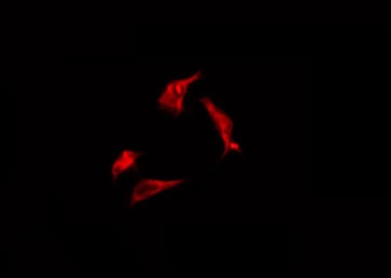
DF7576 staining A549 cells by ICC/IF. Cells were fixed with PFA and permeabilized in 0.1% saponin prior to blocking in 10% serum for 45 minutes at 37!aC. The primary antibody was diluted 1/200 and incubated with the sample for 1 hour at 37!aC. A Alexa Fluor 594 conjugated goat polyclonal to rabbit IgG (H+L), diluted 1/600 was used as secondary antibod
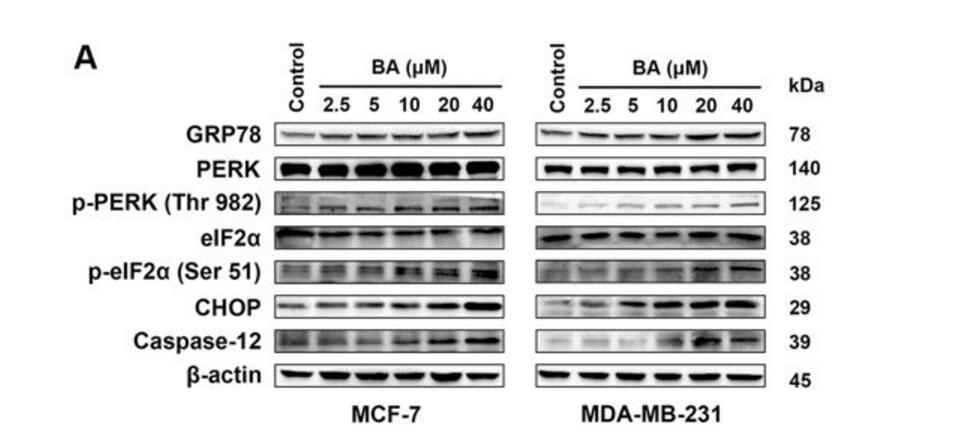
MCF-7 and MDA-MB-231 cells were treated with the indicated concentrations of BA for 24?h, and the protein levels of ER stress-associated signals were stimulated by BA in a dose-dependent manner, including GRP78, p-PERK/PERK, p-eIF2ƒ¿/eIF2ƒ¿, CHOP, and caspase-12.
DF7576 staining Hela by IF/ICC. The sample were fixed with PFA and permeabilized in 0.1% Triton X-100,then blocked in 10% serum for 45 minutes at 25!aC. The primary antibody was diluted at 1/200 and incubated with the sample for 1 hour at 37!aC. An Alexa Fluor 594 conjugated goat anti-rabbit IgG (H+L) Ab, diluted at 1/600, was used as the secondary antibod