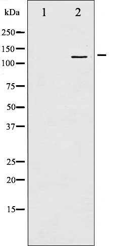
Western blot analysis of FAK phosphorylation expression in EGF treated 293 whole cell lysates,The lane on the left is treated with the antigen-specific peptide.
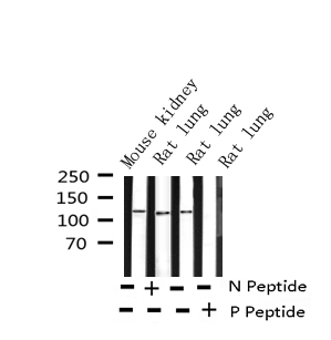
Western blot analysis of Phospho-FAK (Tyr397) expression in various lysates
AF3398 at 1/200 staining Human heart tissue sections by IHC-P. The tissue was formaldehyde fixed and a heat mediated antigen retrieval step in citrate buffer was performed. The tissue was then blocked and incubated with the antibody for 1.5 hours at 22!aC. An HRP conjugated goat anti-rabbit antibody was used as the secondary
AF3398 at 1/200 staining Human heart tissue sections by IHC-P. The tissue was formaldehyde fixed and a heat mediated antigen retrieval step in citrate buffer was performed. The tissue was then blocked and incubated with the antibody for 1.5 hours at 22!aC. An HRP conjugated goat anti-rabbit antibody was used as the secondary

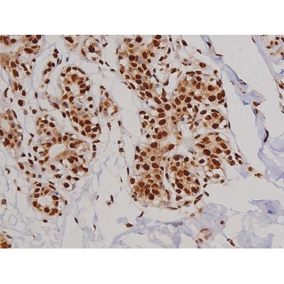
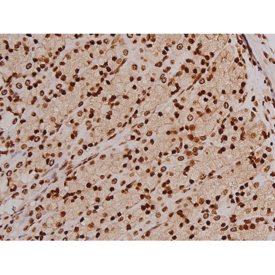
AF3398 at 1/200 staining Human lung cancer tissue sections by IHC-P. The tissue was formaldehyde fixed and a heat mediated antigen retrieval step in citrate buffer was performed. The tissue was then blocked and incubated with the antibody for 1.5 hours at 22!aC. An HRP conjugated goat anti-rabbit antibody was used as the secondary

AF3398 at 1/200 staining Human lung cancer tissue sections by IHC-P. The tissue was formaldehyde fixed and a heat mediated antigen retrieval step in citrate buffer was performed. The tissue was then blocked and incubated with the antibody for 1.5 hours at 22!aC. An HRP conjugated goat anti-rabbit antibody was used as the secondary
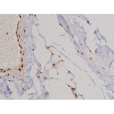
AF3398 at 1/200 staining Human esophagus tissue sections by IHC-P. The tissue was formaldehyde fixed and a heat mediated antigen retrieval step in citrate buffer was performed. The tissue was then blocked and incubated with the antibody for 1.5 hours at 22!aC. An HRP conjugated goat anti-rabbit antibody was used as the secondary
AF3398 at 1/200 staining Human esophagus tissue sections by IHC-P. The tissue was formaldehyde fixed and a heat mediated antigen retrieval step in citrate buffer was performed. The tissue was then blocked and incubated with the antibody for 1.5 hours at 22!aC. An HRP conjugated goat anti-rabbit antibody was used as the secondary
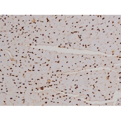
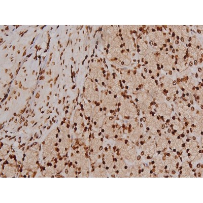
AF3398 at 1/200 staining Mouse heart tissue sections by IHC-P. The tissue was formaldehyde fixed and a heat mediated antigen retrieval step in citrate buffer was performed. The tissue was then blocked and incubated with the antibody for 1.5 hours at 22!aC. An HRP conjugated goat anti-rabbit antibody was used as the secondary
AF3398 at 1/200 staining Mouse heart tissue sections by IHC-P. The tissue was formaldehyde fixed and a heat mediated antigen retrieval step in citrate buffer was performed. The tissue was then blocked and incubated with the antibody for 1.5 hours at 22!aC. An HRP conjugated goat anti-rabbit antibody was used as the secondary
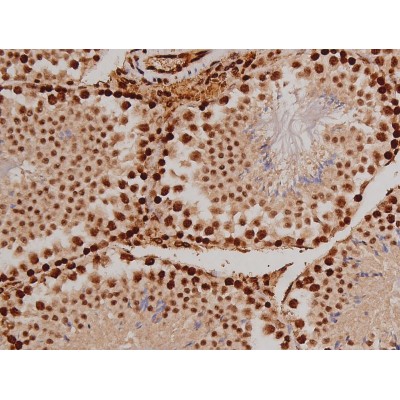
AF3398 at 1/200 staining Mouse testis tissue sections by IHC-P. The tissue was formaldehyde fixed and a heat mediated antigen retrieval step in citrate buffer was performed. The tissue was then blocked and incubated with the antibody for 1.5 hours at 22!aC. An HRP conjugated goat anti-rabbit antibody was used as the secondary
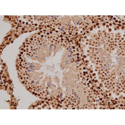
AF3398 at 1/200 staining Mouse testis tissue sections by IHC-P. The tissue was formaldehyde fixed and a heat mediated antigen retrieval step in citrate buffer was performed. The tissue was then blocked and incubated with the antibody for 1.5 hours at 22!aC. An HRP conjugated goat anti-rabbit antibody was used as the secondary
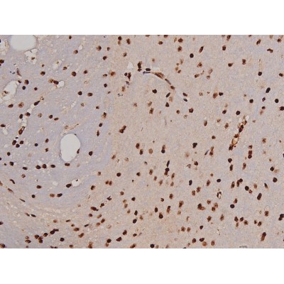
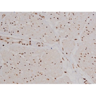
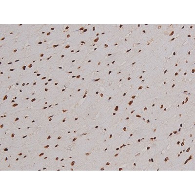
AF3398 at 1/200 staining Mouse brain tissue sections by IHC-P. The tissue was formaldehyde fixed and a heat mediated antigen retrieval step in citrate buffer was performed. The tissue was then blocked and incubated with the antibody for 1.5 hours at 22!aC. An HRP conjugated goat anti-rabbit antibody was used as the secondary
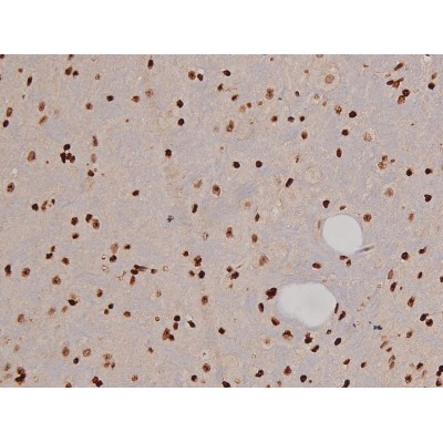
AF3398 at 1/200 staining Mouse brain tissue sections by IHC-P. The tissue was formaldehyde fixed and a heat mediated antigen retrieval step in citrate buffer was performed. The tissue was then blocked and incubated with the antibody for 1.5 hours at 22!aC. An HRP conjugated goat anti-rabbit antibody was used as the secondary

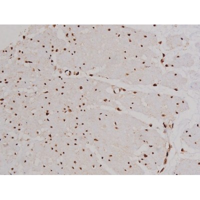
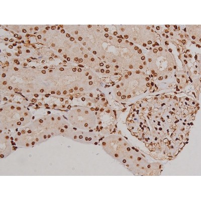
AF3398 at 1/200 staining Rat heart tissue sections by IHC-P. The tissue was formaldehyde fixed and a heat mediated antigen retrieval step in citrate buffer was performed. The tissue was then blocked and incubated with the antibody for 1.5 hours at 22!aC. An HRP conjugated goat anti-rabbit antibody was used as the secondary
AF3398 at 1/200 staining Rat heart tissue sections by IHC-P. The tissue was formaldehyde fixed and a heat mediated antigen retrieval step in citrate buffer was performed. The tissue was then blocked and incubated with the antibody for 1.5 hours at 22!aC. An HRP conjugated goat anti-rabbit antibody was used as the secondary
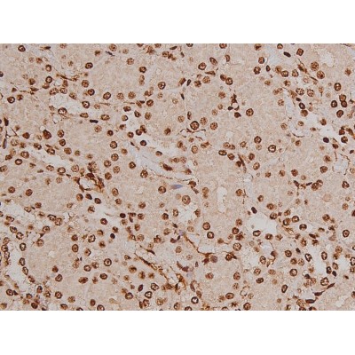
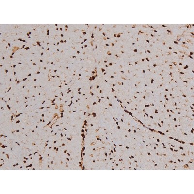
AF3398 at 1/200 staining Rat kidney tissue sections by IHC-P. The tissue was formaldehyde fixed and a heat mediated antigen retrieval step in citrate buffer was performed. The tissue was then blocked and incubated with the antibody for 1.5 hours at 22!aC. An HRP conjugated goat anti-rabbit antibody was used as the secondary
AF3398 at 1/200 staining Rat kidney tissue sections by IHC-P. The tissue was formaldehyde fixed and a heat mediated antigen retrieval step in citrate buffer was performed. The tissue was then blocked and incubated with the antibody for 1.5 hours at 22!aC. An HRP conjugated goat anti-rabbit antibody was used as the secondary
AF3398 at 1/200 staining Rat ganstric tissue sections by IHC-P. The tissue was formaldehyde fixed and a heat mediated antigen retrieval step in citrate buffer was performed. The tissue was then blocked and incubated with the antibody for 1.5 hours at 22!aC. An HRP conjugated goat anti-rabbit antibody was used as the secondary
AF3398 at 1/200 staining Rat ganstric tissue sections by IHC-P. The tissue was formaldehyde fixed and a heat mediated antigen retrieval step in citrate buffer was performed. The tissue was then blocked and incubated with the antibody for 1.5 hours at 22!aC. An HRP conjugated goat anti-rabbit antibody was used as the secondary