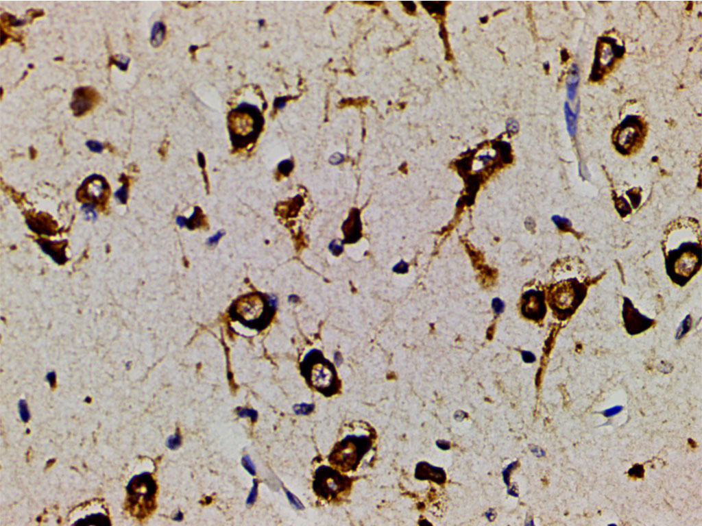
AF3241 at 1/200 staining human brain tissue sections by IHC-P. The tissue was formaldehyde fixed and a heat mediated antigen retrieval step in citrate buffer was performed. The tissue was then blocked and incubated with the antibody for 1.5 hours at 22!aC. An HRP conjugated goat anti-rabbit antibody was used as the secondary
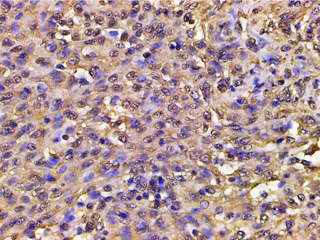
AF3241 at 1/200 staining human meningeal carcinomatosis(MC) tissue sections by IHC-P. The tissue was formaldehyde fixed and a heat mediated antigen retrieval step in citrate buffer was performed. The tissue was then blocked and incubated with the antibody for 1.5 hours at 22!aC. An HRP conjugated goat anti-rabbit antibody was used as the secondary
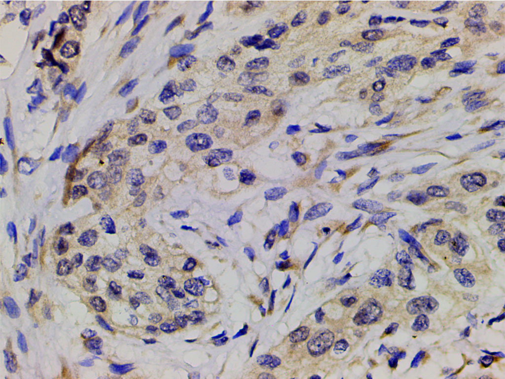
AF3241 at 1/200 staining human esophageal carcinoma tissue sections by IHC-P. The tissue was formaldehyde fixed and a heat mediated antigen retrieval step in citrate buffer was performed. The tissue was then blocked and incubated with the antibody for 1.5 hours at 22!aC. An HRP conjugated goat anti-rabbit antibody was used as the secondary
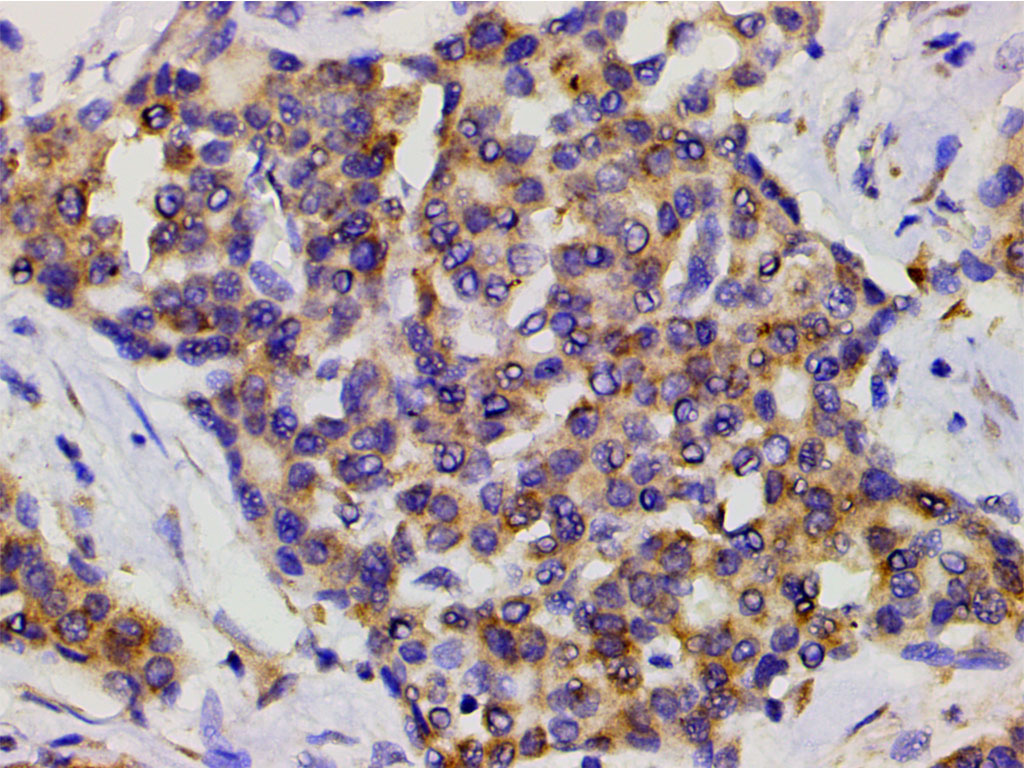
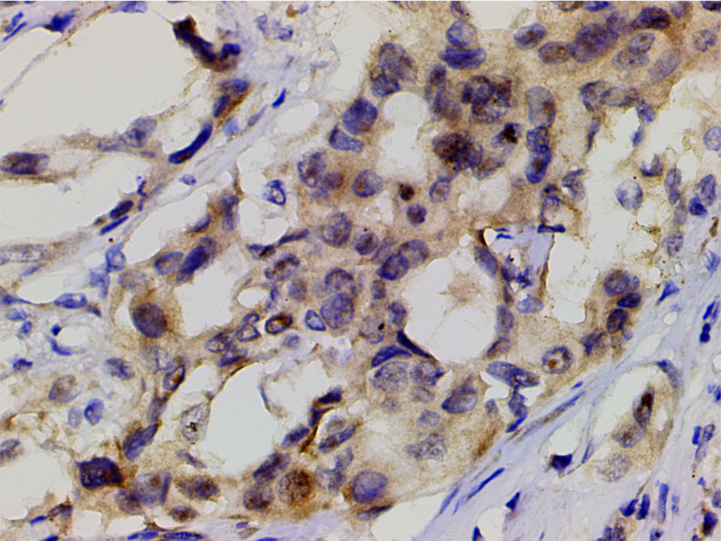
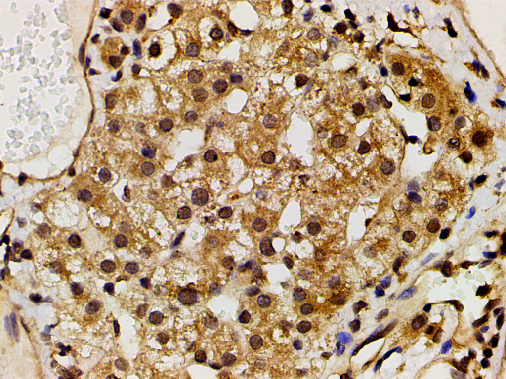
AF3241 at 1/200 staining human liver cancer tissue sections by IHC-P. The tissue was formaldehyde fixed and a heat mediated antigen retrieval step in citrate buffer was performed. The tissue was then blocked and incubated with the antibody for 1.5 hours at 22!aC. An HRP conjugated goat anti-rabbit antibody was used as the secondary
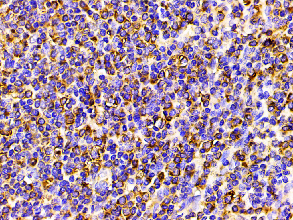
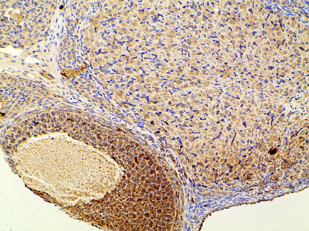
AF3241 at 1/200 staining human spleen tissue sections by IHC-P. The tissue was formaldehyde fixed and a heat mediated antigen retrieval step in citrate buffer was performed. The tissue was then blocked and incubated with the antibody for 1.5 hours at 22!aC. An HRP conjugated goat anti-rabbit antibody was used as the secondary
AF3241 at 1/200 staining human lung cancer tissue sections by IHC-P. The tissue was formaldehyde fixed and a heat mediated antigen retrieval step in citrate buffer was performed. The tissue was then blocked and incubated with the antibody for 1.5 hours at 22!aC. An HRP conjugated goat anti-rabbit antibody was used as the secondary
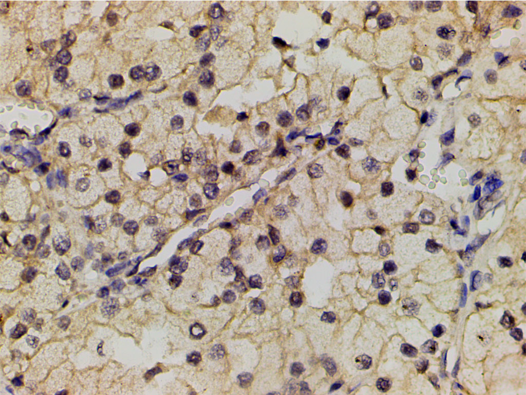
AF3241 at 1/200 staining human renal clear cell carcinoma tissue sections by IHC-P. The tissue was formaldehyde fixed and a heat mediated antigen retrieval step in citrate buffer was performed. The tissue was then blocked and incubated with the antibody for 1.5 hours at 22!aC. An HRP conjugated goat anti-rabbit antibody was used as the secondary
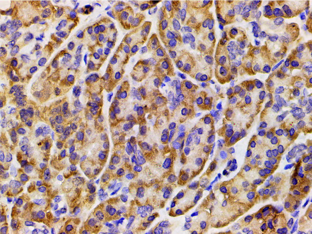
AF3241 at 1/200 staining human colon cancer tissue sections by IHC-P. The tissue was formaldehyde fixed and a heat mediated antigen retrieval step in citrate buffer was performed. The tissue was then blocked and incubated with the antibody for 1.5 hours at 22!aC. An HRP conjugated goat anti-rabbit antibody was used as the secondary
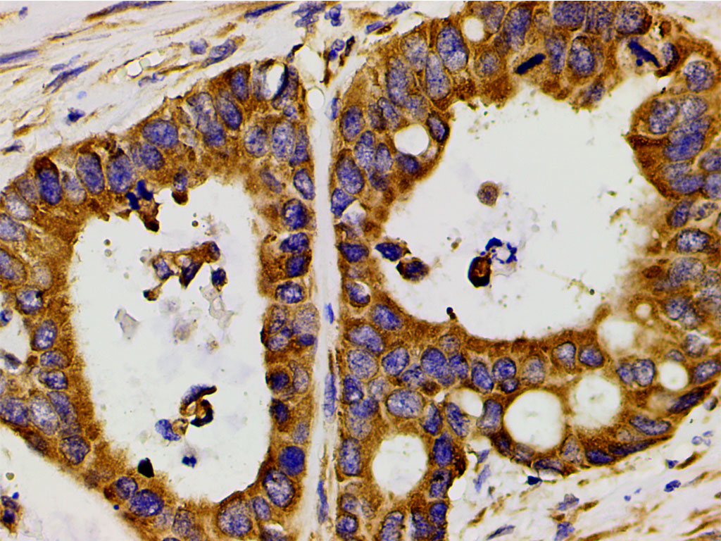
AF3241 at 1/200 staining human duodenum tissue sections by IHC-P. The tissue was formaldehyde fixed and a heat mediated antigen retrieval step in citrate buffer was performed. The tissue was then blocked and incubated with the antibody for 1.5 hours at 22!aC. An HRP conjugated goat anti-rabbit antibody was used as the secondary

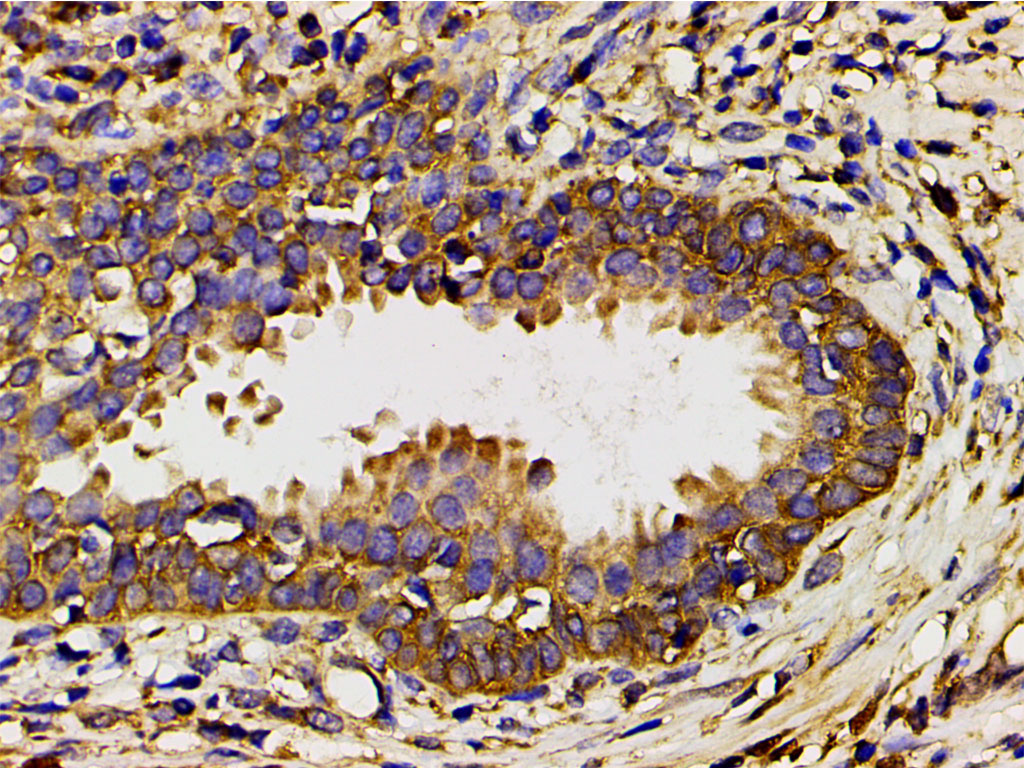
AF3241 at 1/200 staining human bladder cancer tissue sections by IHC-P. The tissue was formaldehyde fixed and a heat mediated antigen retrieval step in citrate buffer was performed. The tissue was then blocked and incubated with the antibody for 1.5 hours at 22!aC. An HRP conjugated goat anti-rabbit antibody was used as the secondary

AF3241 at 1/200 staining human placenta tissue sections by IHC-P. The tissue was formaldehyde fixed and a heat mediated antigen retrieval step in citrate buffer was performed. The tissue was then blocked and incubated with the antibody for 1.5 hours at 22!aC. An HRP conjugated goat anti-rabbit antibody was used as the secondary
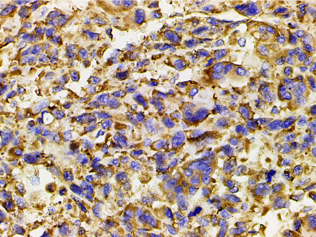
AF3241 at 1/200 staining human vascular cancer tissue sections by IHC-P. The tissue was formaldehyde fixed and a heat mediated antigen retrieval step in citrate buffer was performed. The tissue was then blocked and incubated with the antibody for 1.5 hours at 22!aC. An HRP conjugated goat anti-rabbit antibody was used as the secondary

AF3241 at 1/200 staining human seminoma tissue sections by IHC-P. The tissue was formaldehyde fixed and a heat mediated antigen retrieval step in citrate buffer was performed. The tissue was then blocked and incubated with the antibody for 1.5 hours at 22!aC. An HRP conjugated goat anti-rabbit antibody was used as the secondary
AF3241 at 1/200 staining human myosarcoma tissue sections by IHC-P. The tissue was formaldehyde fixed and a heat mediated antigen retrieval step in citrate buffer was performed. The tissue was then blocked and incubated with the antibody for 1.5 hours at 22!aC. An HRP conjugated goat anti-rabbit antibody was used as the secondary
AF3241 at 1/200 staining human osteosarcoma tissue sections by IHC-P. The tissue was formaldehyde fixed and a heat mediated antigen retrieval step in citrate buffer was performed. The tissue was then blocked and incubated with the antibody for 1.5 hours at 22!aC. An HRP conjugated goat anti-rabbit antibody was used as the secondary
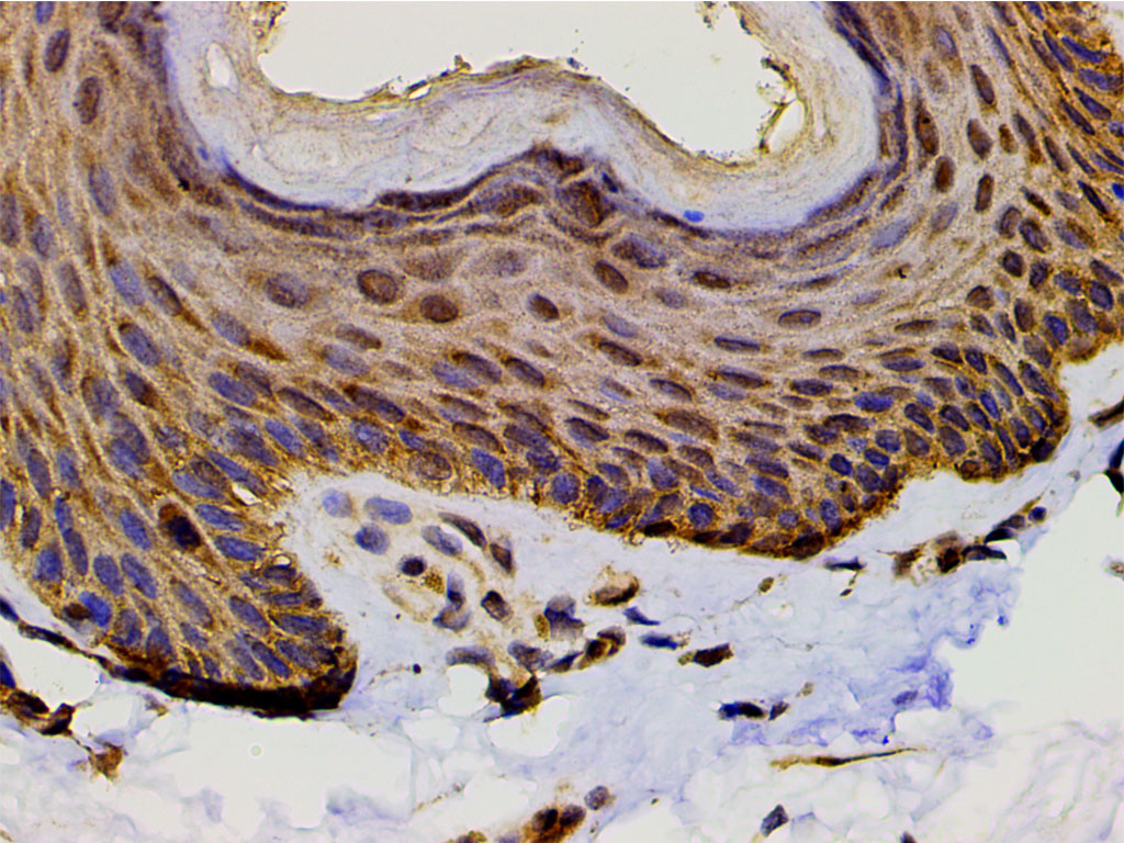
AF3241 at 1/200 staining human skin tissue sections by IHC-P. The tissue was formaldehyde fixed and a heat mediated antigen retrieval step in citrate buffer was performed. The tissue was then blocked and incubated with the antibody for 1.5 hours at 22!aC. An HRP conjugated goat anti-rabbit antibody was used as the secondary
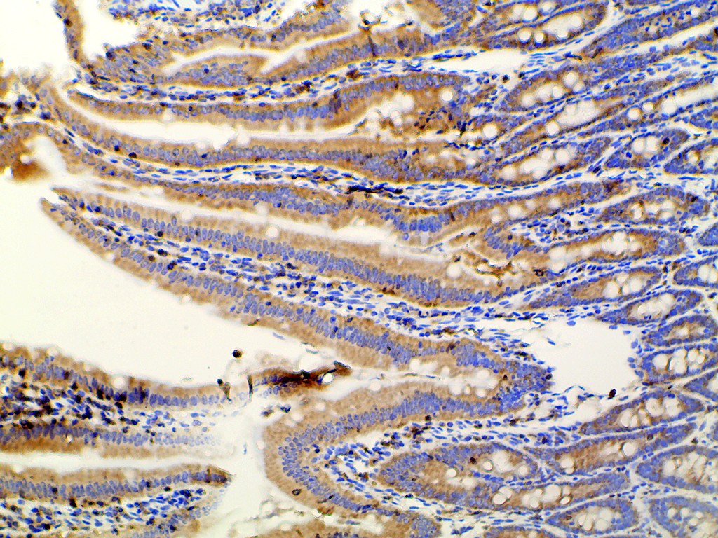
AF3241 at 1/100 staining rat Intestinal tissue sections by IHC-P. The tissue was formaldehyde fixed and a heat mediated antigen retrieval step in citrate buffer was performed. The tissue was then blocked and incubated with the antibody for 1.5 hours at 22!aC. An HRP conjugated goat anti-rabbit antibody was used as the secondary
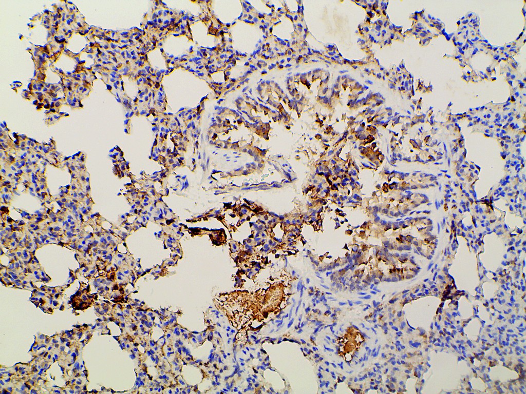
AF3241 at 1/100 staining rat lung tissue sections by IHC-P. The tissue was formaldehyde fixed and a heat mediated antigen retrieval step in citrate buffer was performed. The tissue was then blocked and incubated with the antibody for 1.5 hours at 22!aC. An HRP conjugated goat anti-rabbit antibody was used as the secondary
AF3241 at 1/100 staining rat ovarian tissue sections by IHC-P. The tissue was formaldehyde fixed and a heat mediated antigen retrieval step in citrate buffer was performed. The tissue was then blocked and incubated with the antibody for 1.5 hours at 22!aC. An HRP conjugated goat anti-rabbit antibody was used as the secondary
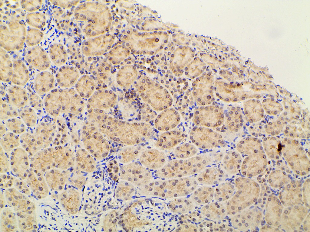
AF3241 at 1/100 staining rat kidney tissue sections by IHC-P. The tissue was formaldehyde fixed and a heat mediated antigen retrieval step in citrate buffer was performed. The tissue was then blocked and incubated with the antibody for 1.5 hours at 22!aC. An HRP conjugated goat anti-rabbit antibody was used as the secondary