Western blot analysis of eIF2 alpha phosphorylation expression in IFN-ƒ¿ treated K562 whole cell lysates,The lane on the left is treated with the antigen-specific peptide.
AF3087 at 1/100 staining rat ovarian tissue sections by IHC-P. The tissue was formaldehyde fixed and a heat mediated antigen retrieval step in citrate buffer was performed. The tissue was then blocked and incubated with the antibody for 1.5 hours at 22!aC. An HRP conjugated goat anti-rabbit antibody was used as the secondary
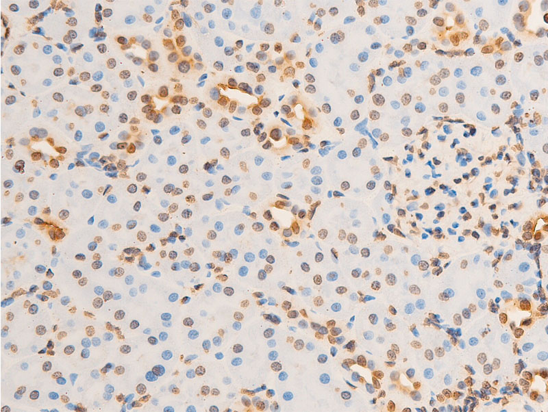
AF3087 at 1/100 staining rat kidney tissue sections by IHC-P. The tissue was formaldehyde fixed and a heat mediated antigen retrieval step in citrate buffer was performed. The tissue was then blocked and incubated with the antibody for 1.5 hours at 22!aC. An HRP conjugated goat anti-rabbit antibody was used as the secondary
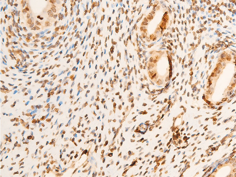
AF3087 at 1/100 staining rat uterine tissue sections by IHC-P. The tissue was formaldehyde fixed and a heat mediated antigen retrieval step in citrate buffer was performed. The tissue was then blocked and incubated with the antibody for 1.5 hours at 22!aC. An HRP conjugated goat anti-rabbit antibody was used as the secondary
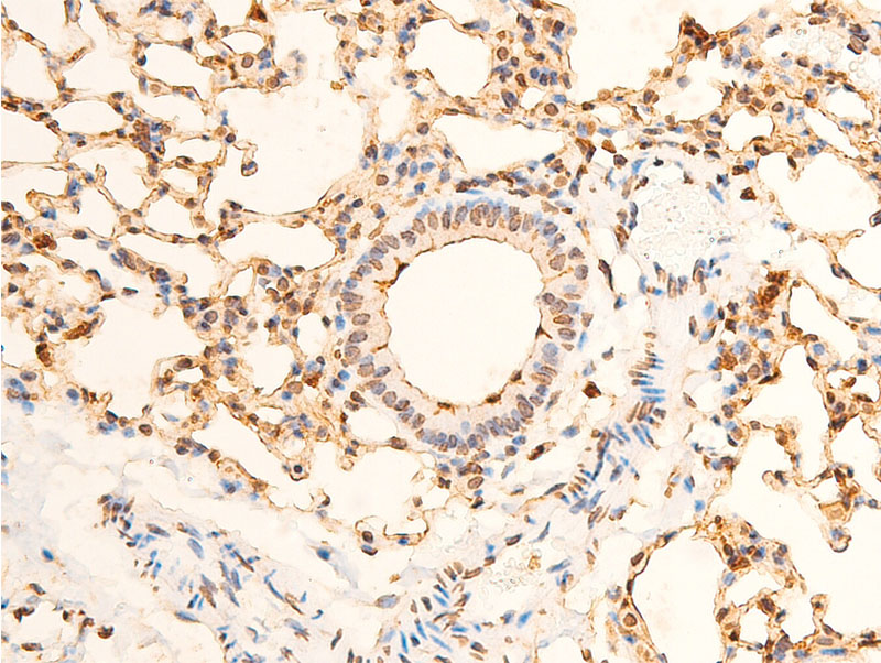
AF3087 at 1/100 staining mouse lung tissue sections by IHC-P. The tissue was formaldehyde fixed and a heat mediated antigen retrieval step in citrate buffer was performed. The tissue was then blocked and incubated with the antibody for 1.5 hours at 22!aC. An HRP conjugated goat anti-rabbit antibody was used as the secondary
AF3087 at 1/100 staining mouse kidney tissue sections by IHC-P. The tissue was formaldehyde fixed and a heat mediated antigen retrieval step in citrate buffer was performed. The tissue was then blocked and incubated with the antibody for 1.5 hours at 22!aC. An HRP conjugated goat anti-rabbit antibody was used as the secondary
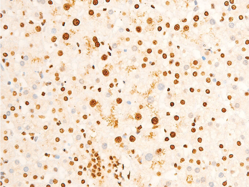
AF3087 at 1/100 staining mouse gastric tissue sections by IHC-P. The tissue was formaldehyde fixed and a heat mediated antigen retrieval step in citrate buffer was performed. The tissue was then blocked and incubated with the antibody for 1.5 hours at 22!aC. An HRP conjugated goat anti-rabbit antibody was used as the secondary
AF3087 at 1/100 staining human lung tissue sections by IHC-P. The tissue was formaldehyde fixed and a heat mediated antigen retrieval step in citrate buffer was performed. The tissue was then blocked and incubated with the antibody for 1.5 hours at 22!aC. An HRP conjugated goat anti-rabbit antibody was used as the secondary
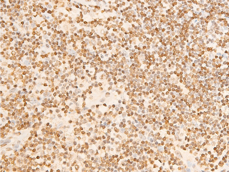
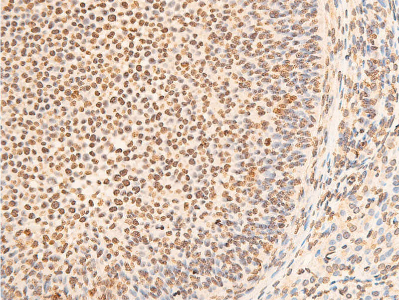
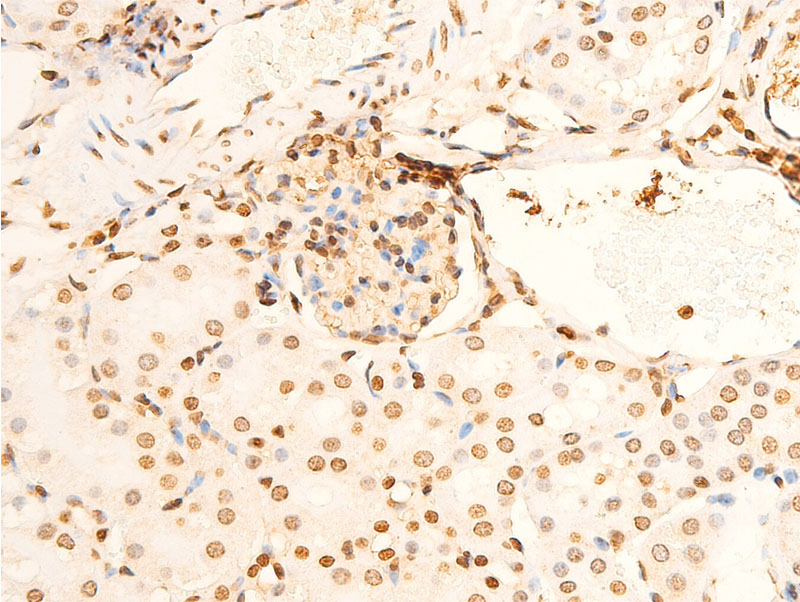
AF3087 at 1/100 staining human appendiceal tissue sections by IHC-P. The tissue was formaldehyde fixed and a heat mediated antigen retrieval step in citrate buffer was performed. The tissue was then blocked and incubated with the antibody for 1.5 hours at 22!aC. An HRP conjugated goat anti-rabbit antibody was used as the secondary
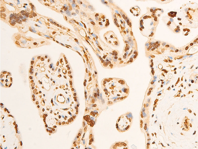
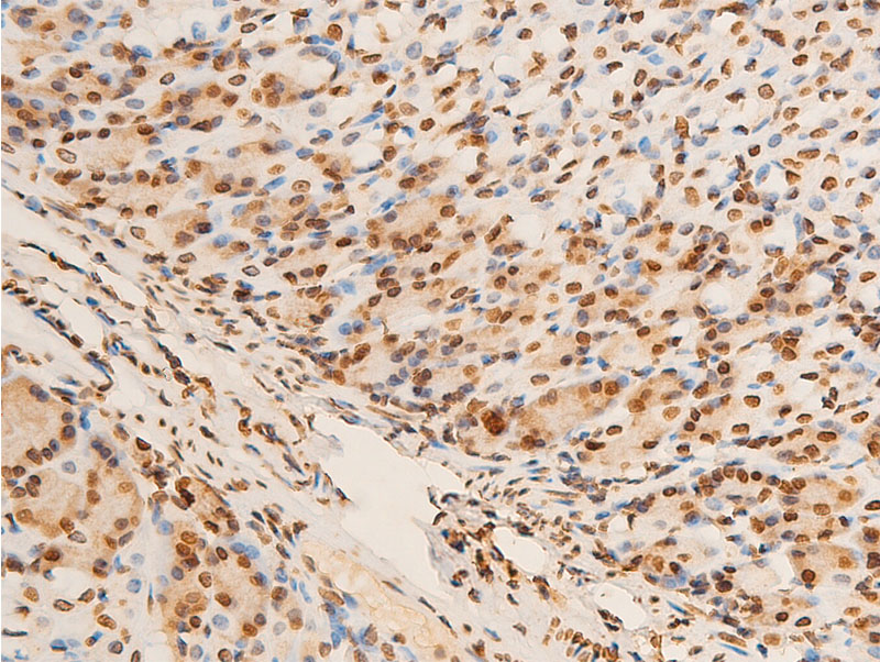
AF3087 at 1/100 staining human placenta tissue sections by IHC-P. The tissue was formaldehyde fixed and a heat mediated antigen retrieval step in citrate buffer was performed. The tissue was then blocked and incubated with the antibody for 1.5 hours at 22!aC. An HRP conjugated goat anti-rabbit antibody was used as the secondary
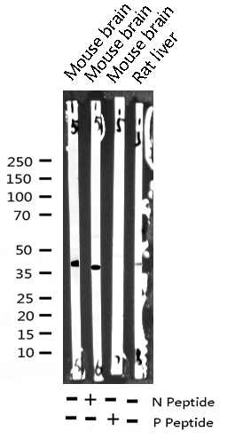
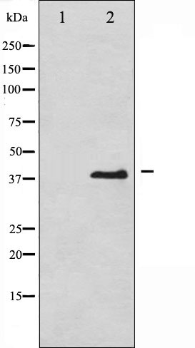
Western blot analysis of Phospho-eIF2 alpha (Ser51) expression in various lysates
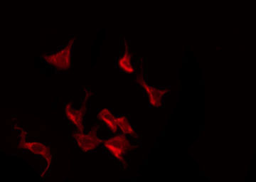
AF3087 staining K562 by IF/ICC. The sample were fixed with PFA and permeabilized in 0.1% Triton X-100,then blocked in 10% serum for 45 minutes at 25!aC. The primary antibody was diluted at 1/200 and incubated with the sample for 1 hour at 37!aC. An Alexa Fluor 594 conjugated goat anti-rabbit IgG (H+L) Ab, diluted at 1/600, was used as the secondary antibod