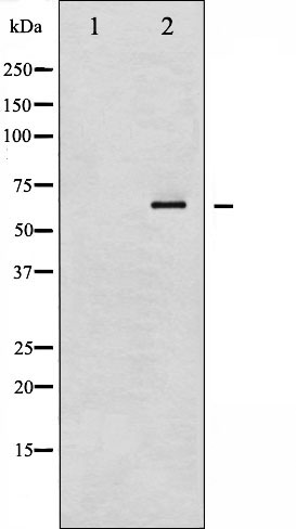
Western blot analysis of NF kappaB p65 phosphorylation expression in IL-1 treated Raw264.7 whole cell lysates,The lane on the left is treated with the antigen-specific peptide.
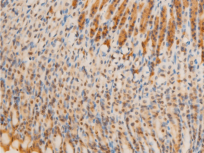
AF2006 at 1/100 staining mouse gastric tissue sections by IHC-P. The tissue was formaldehyde fixed and a heat mediated antigen retrieval step in citrate buffer was performed. The tissue was then blocked and incubated with the antibody for 1.5 hours at 22!aC. An HRP conjugated goat anti-rabbit antibody was used as the secondary
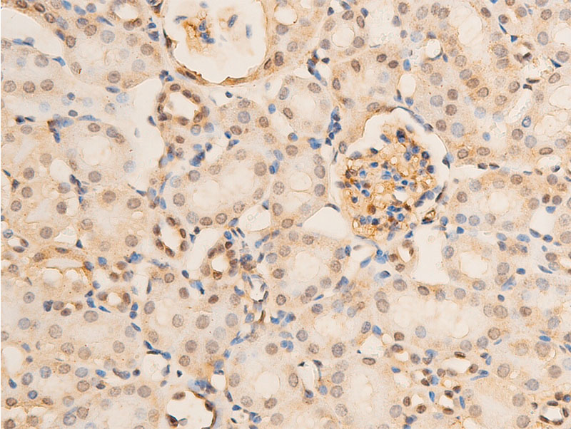
AF2006 at 1/100 staining mouse kidney tissue sections by IHC-P. The tissue was formaldehyde fixed and a heat mediated antigen retrieval step in citrate buffer was performed. The tissue was then blocked and incubated with the antibody for 1.5 hours at 22!aC. An HRP conjugated goat anti-rabbit antibody was used as the secondary
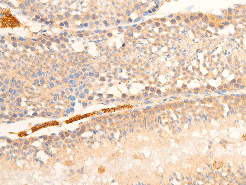
AF2006 at 1/100 staining mouse testis tissue sections by IHC-P. The tissue was formaldehyde fixed and a heat mediated antigen retrieval step in citrate buffer was performed. The tissue was then blocked and incubated with the antibody for 1.5 hours at 22!aC. An HRP conjugated goat anti-rabbit antibody was used as the secondary
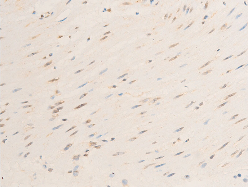
AF2006 at 1/100 staining human heart tissue sections by IHC-P. The tissue was formaldehyde fixed and a heat mediated antigen retrieval step in citrate buffer was performed. The tissue was then blocked and incubated with the antibody for 1.5 hours at 22!aC. An HRP conjugated goat anti-rabbit antibody was used as the secondary
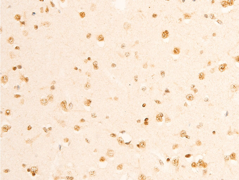
AF2006 at 1/100 staining human brain tissue sections by IHC-P. The tissue was formaldehyde fixed and a heat mediated antigen retrieval step in citrate buffer was performed. The tissue was then blocked and incubated with the antibody for 1.5 hours at 22!aC. An HRP conjugated goat anti-rabbit antibody was used as the secondary
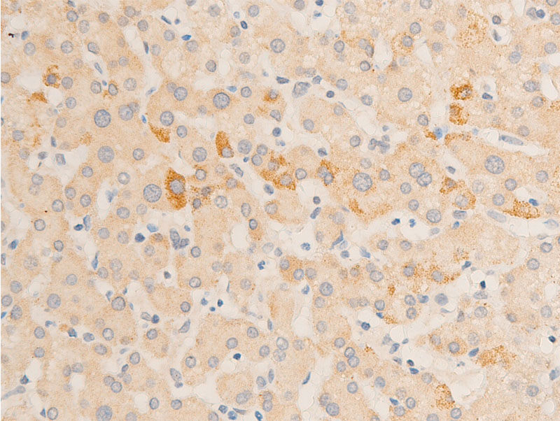
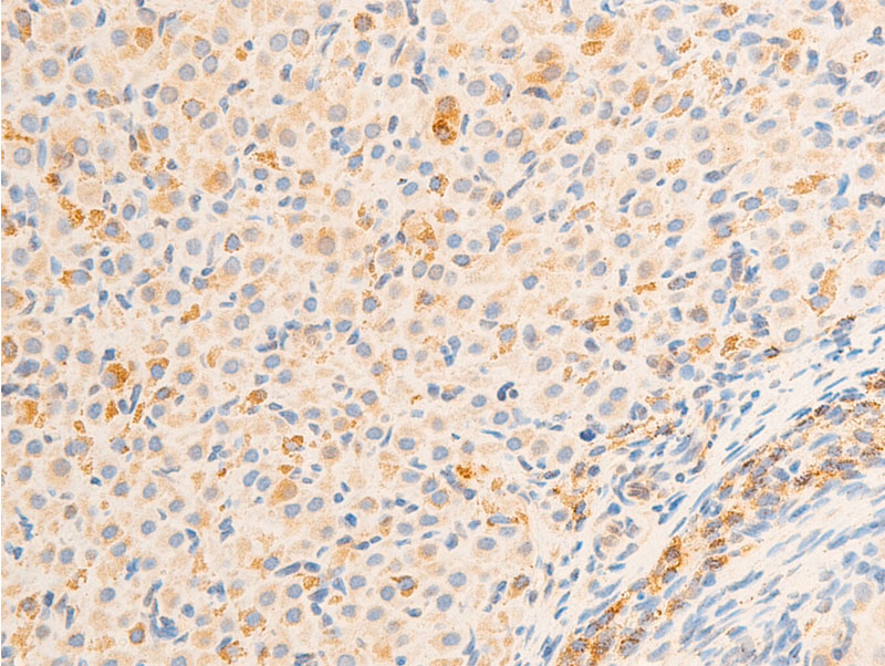
AF2006 at 1/100 staining human liver tissue sections by IHC-P. The tissue was formaldehyde fixed and a heat mediated antigen retrieval step in citrate buffer was performed. The tissue was then blocked and incubated with the antibody for 1.5 hours at 22!aC. An HRP conjugated goat anti-rabbit antibody was used as the secondary
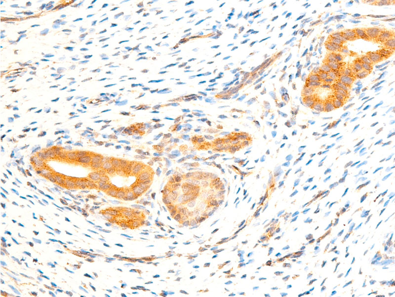
AF2006 at 1/100 staining rat uterine tissue sections by IHC-P. The tissue was formaldehyde fixed and a heat mediated antigen retrieval step in citrate buffer was performed. The tissue was then blocked and incubated with the antibody for 1.5 hours at 22!aC. An HRP conjugated goat anti-rabbit antibody was used as the secondary
AF2006 at 1/100 staining rat ovarian tissue sections by IHC-P. The tissue was formaldehyde fixed and a heat mediated antigen retrieval step in citrate buffer was performed. The tissue was then blocked and incubated with the antibody for 1.5 hours at 22!aC. An HRP conjugated goat anti-rabbit antibody was used as the secondary
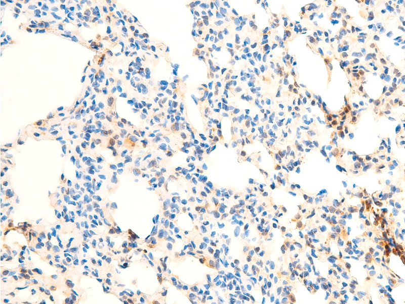
AF2006 at 1/100 staining rat lung tissue sections by IHC-P. The tissue was formaldehyde fixed and a heat mediated antigen retrieval step in citrate buffer was performed. The tissue was then blocked and incubated with the antibody for 1.5 hours at 22!aC. An HRP conjugated goat anti-rabbit antibody was used as the secondary
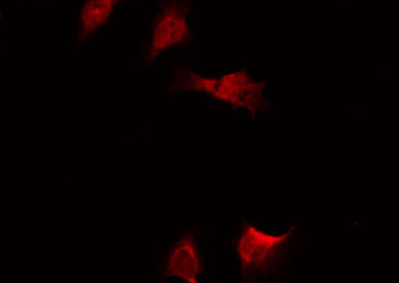
AF2006 staining lovo cells by ICC/IF. Cells were fixed with PFA and permeabilized in 0.1% saponin prior to blocking in 10% serum for 45 minutes at 37!aC. The primary antibody was diluted 1/400 and incubated with the sample for 1 hour at 37!aC. A Alexa Fluor? 594 conjugated goat polyclonal to rabbit IgG (H+L), diluted 1/600 was used as secondary antibod
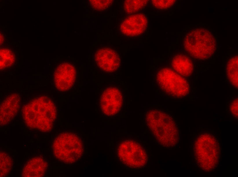
AF2006 staining HeLa by IF/ICC. The sample were fixed with PFA and permeabilized in 0.1% Triton X-100,then blocked in 10% serum for 45 minutes at 25!aC. The primary antibody was diluted at 1/200 and incubated with the sample for 1 hour at 37!aC. An Alexa Fluor 594 conjugated goat anti-rabbit IgG (H+L) Ab, diluted at 1/600, was used as the secondary antibod