Western blot analysis on 293 cell lysate using E-cadherin Antibody,The lane on the left is treated with the antigen-specific peptide.
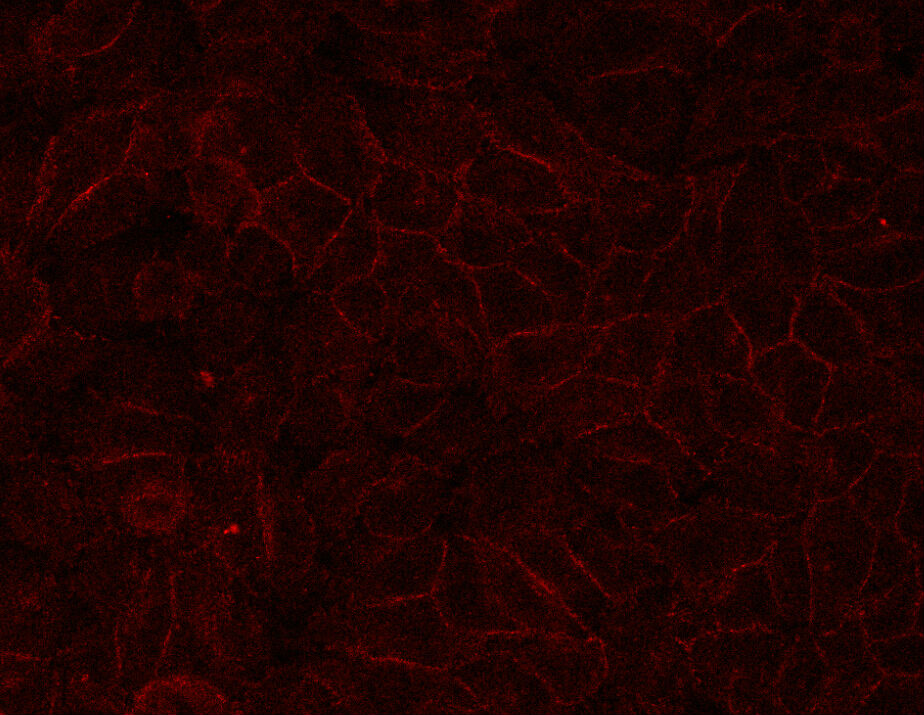
E-cadherin for IHC in human HepG2,Provided by Tianjin University
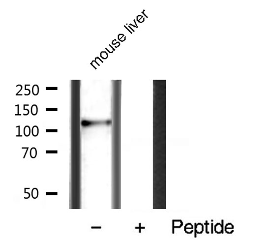
Western blot analysis on mouse liver tissue lysate using E-cadherin Antibody
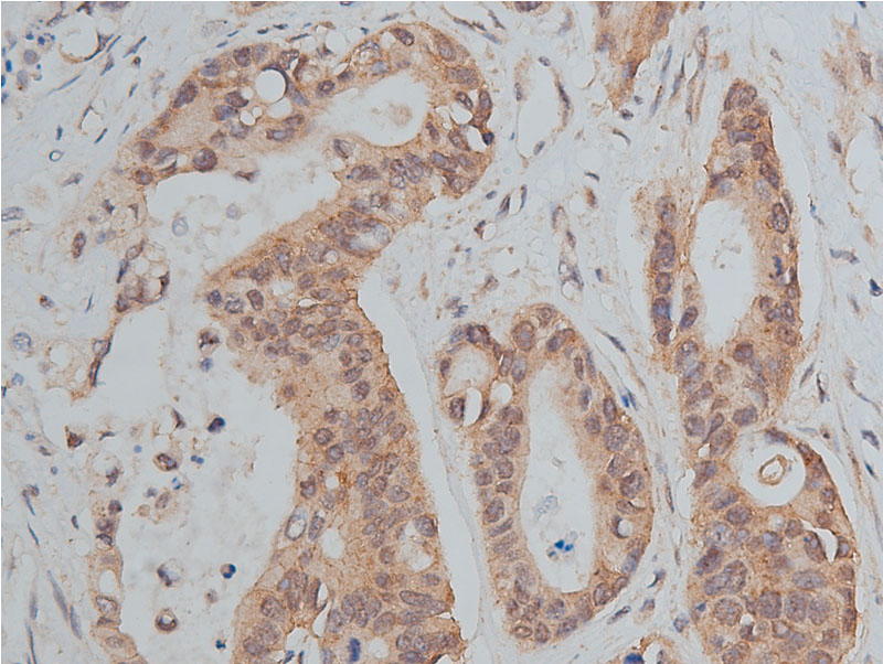
AF0131 at 1/50 staining human colon cancer tissue sections by IHC-P. The tissue was formaldehyde fixed and a heat mediated antigen retrieval step in citrate buffer was performed. The tissue was then blocked and incubated with the antibody for 1.5 hours at 22!aC. An HRP conjugated goat anti-rabbit antibody was used as the secondary
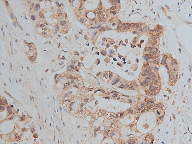
AF0131 at 1/50 staining human colon cancer tissue sections by IHC-P. The tissue was formaldehyde fixed and a heat mediated antigen retrieval step in citrate buffer was performed. The tissue was then blocked and incubated with the antibody for 1.5 hours at 22!aC. An HRP conjugated goat anti-rabbit antibody was used as the secondary
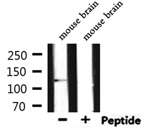
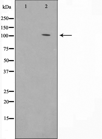
Western blot analysis of extracts from mouse brain, using E-cadherin Antibody.