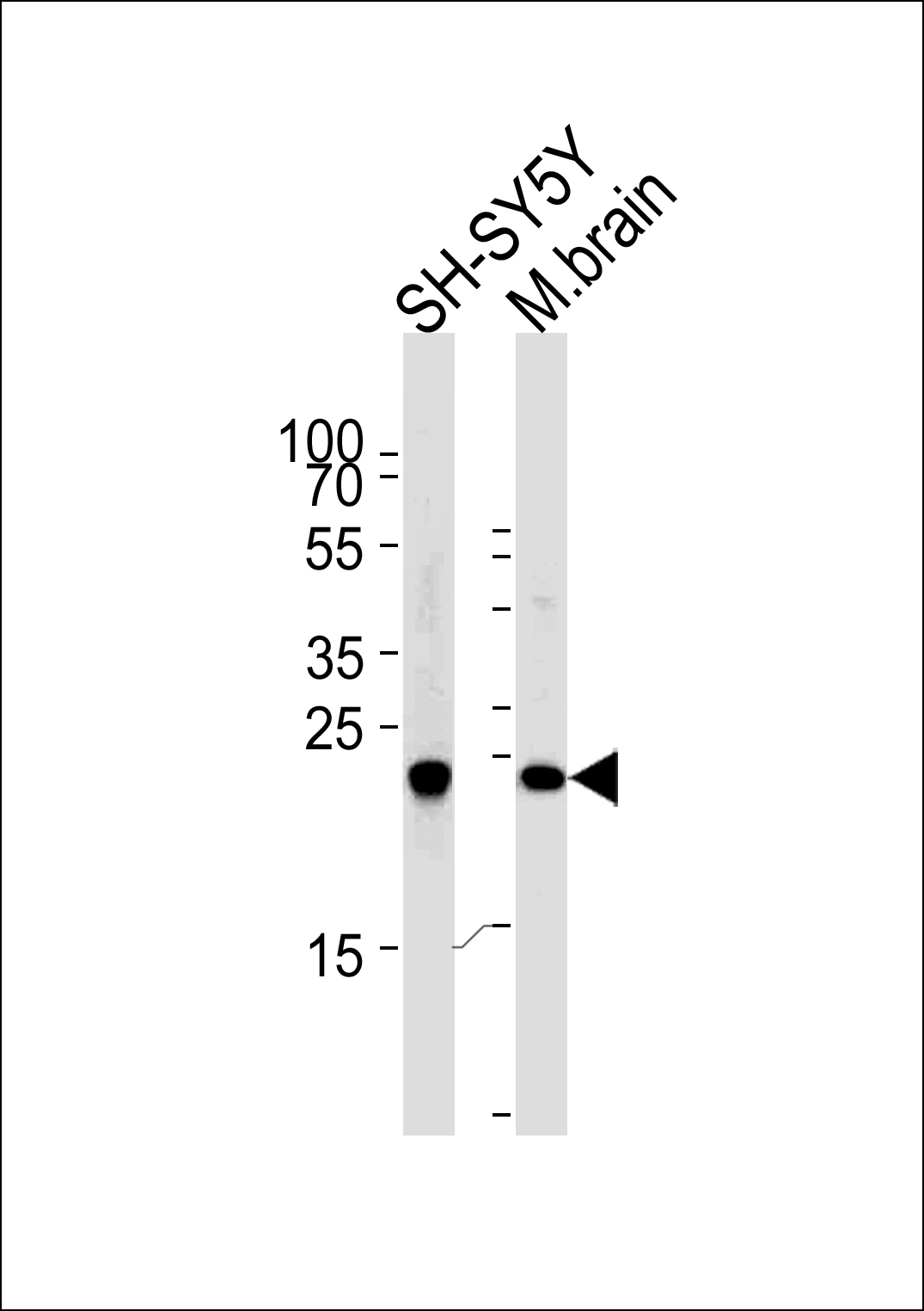
Western blot analysis of lysates from SH-SY5Y cell line and mouse brain tissue (from left to right),using UCHL1 Antibody (C-term)(Cat. #AP2126e).AP2126e was diluted at 1:1000 at each lane. A goat anti-rabbit IgG H&L(HRP) at 1:5000 dilution was used as the secondary antibody.Lysates at 35ug per lane.
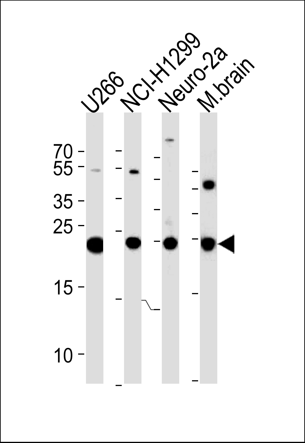
UCHL1 Antibody (C-term) (Cat.# AP2126e) western blot analysis in U266,NCI-H1299,mouse Neuro-2a cell line and mouse brain tissue lysates (35ug/lane).This demonstrates the UCHL1 antibody detected the UCHL1 protein (arrow).

Fluorescent image of U251 cell stained with UCHL1 Antibody (C-term)(Cat#AP2126e/SA120806AG).U251 cells were fixed with 4% PFA (20 min), permeabilized with Triton X-100 (0.1%, 10 min), then incubated with UCHL1 primary antibody (1:25, 1 h at 37Åé). For secondary antibody, Alexa FluorR 488 conjugated donkey anti-rabbit antibody (green) was used (1:400, 50 min at 37Åé).Cytoplasmic actin was counterstained with Alexa FluorR 555 (red) conjugated Phalloidin (7units/ml, 1 h at 37Åé). UCHL1 immunoreactivity is localized to Cytoplasm and Nucleus significantly.
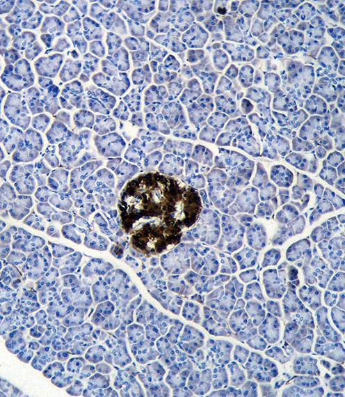
UCHL1 Antibody (C-term) (Cat. #AP2126e)immunohistochemistry analysis in formalin fixed and paraffin embedded human pancreas tissue followed by peroxidase conjugation of the secondary antibody and DAB staining.This data demonstrates the use of UCHL1 Antibody (C-term) for immunohistochemistry. Clinical relevance has not been evaluated.
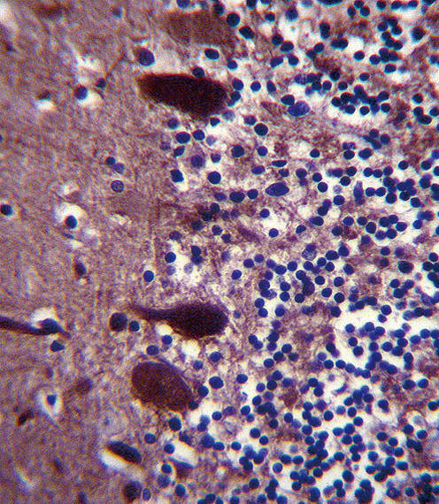
UCHL1 Antibody (C-term) (Cat. #AP2126e)immunohistochemistry analysis in formalin fixed and paraffin embedded human cerebellum tissue followed by peroxidase conjugation of the secondary antibody and DAB staining.This data demonstrates the use of UCHL1 Antibody (C-term) for immunohistochemistry. Clinical relevance has not been evaluated.