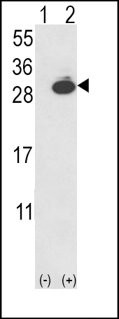
Western blot analysis of UCHL1 (arrow) using rabbit polyclonal UCHL1-V31 (Cat. #AP2126a). 293 cell lysates (2 ug/lane) either nontransfected (Lane 1) or transiently transfected with the UCHL1 gene (Lane 2) (Origene Technologies).
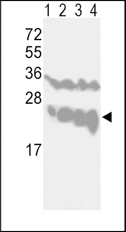
Western blot analysis of UCHL1-V31 (Cat. #AP2126a) in CEM(lane 1), Jurkat(lane 2), Y79(lane 3) cell line and mouse brain tissue(lane 4) lysates (35ug/lane). UCHL1 (arrow) was detected using the purified Pab.
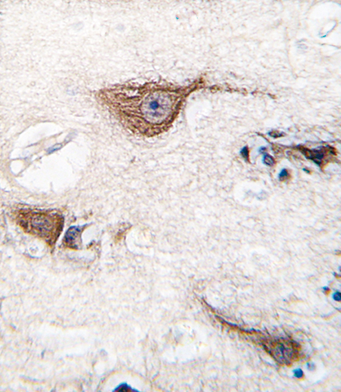
Formalin-fixed and paraffin-embedded human brain tissue reacted with UCHL1 (Park5) antibody (N-term) (Cat.#AP2126a), which was peroxidase-conjugated to the secondary antibody, followed by DAB staining. This data demonstrates the use of this antibody for immunohistochemistry; clinical relevance has not been evaluated.
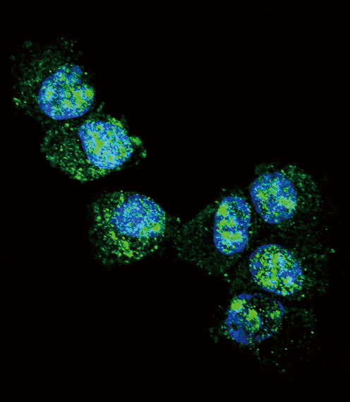
Confocal immunofluorescent analysis of UCHL1 Antibody (N-term)(Cat#AP2126a) with NCI-H460 cell followed by Alexa Fluor 488-conjugated goat anti-rabbit lgG (green).DAPI was used to stain the cell nuclear (blue).
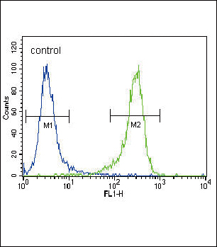
UCHL1 Antibody (N-term) (Cat. #AP2126a) flow cytometric analysis of NCI-H460 cells (right histogram) compared to a negative control cell (left histogram).FITC-conjugated goat-anti-rabbit secondary antibodies were used for the analysis.