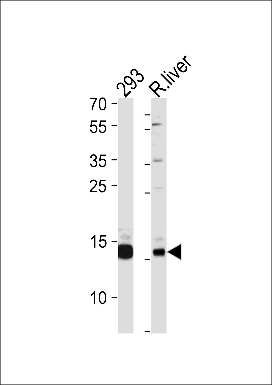
SUMO3 Antibody (Cat. #AP1290a) western blot analysis in 293 cell line and rat liver tissue lysates (35ug/lane).This demonstrates the SUMO3 antibody detected the SUMO3 protein (arrow).
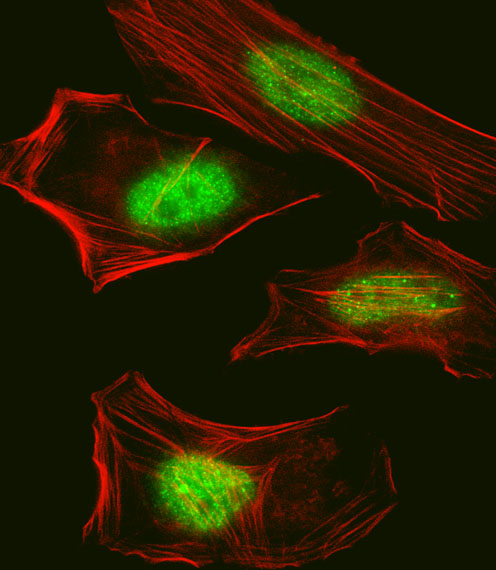
Fluorescent confocal image of Hela cell stained with Pan SUMO Antibody(Cat#AP1290a).Hela cells were fixed with 4% PFA (20 min), permeabilized with Triton X-100 (0.1%, 10 min), then incubated with Pan SUMO primary antibody (1:25, 1 h at 37��). For secondary antibody, Alexa FluorR 488 conjugated donkey anti-rabbit antibody (green) was used (1:400, 50 min at 37��).Cytoplasmic actin was counterstained with Alexa FluorR 555 (red) conjugated Phalloidin (7units/ml, 1 h at 37��). Nuclei were counterstained with DAPI (blue) (10 ��g/ml, 10 min).Pan SUMO immunoreactivity is localized to Nucleus significantly.
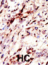
Formalin-fixed and paraffin-embedded human cancer tissue reacted with the primary antibody, which was peroxidase-conjugated to the secondary antibody, followed by AEC staining. This data demonstrates the use of this antibody for immunohistochemistry; clinical relevance has not been evaluated. BC = breast carcinoma; HC = hepatocarcinoma.