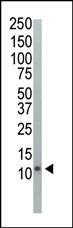
Western blot analysis of anti-Ubiquitin Pab (Cat. #AP1229a) in HeLa cell lysate. Ubiquitin (Arrow) was detected using purified Pab. Secondary HRP-anti-rabbit was used for signal visualization with chemiluminescence.
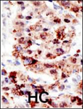
Formalin-fixed and paraffin-embedded human cancer tissue reacted with the primary antibody, which was peroxidase-conjugated to the secondary antibody, followed by AEC staining. This data demonstrates the use of this antibody for immunohistochemistry; clinical relevance has not been evaluated. BC = breast carcinoma; HC = hepatocarcinoma.
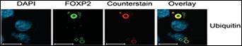
Characterization of FOXP2 Isoforms. FOXP2.10t was detected with an antibody to the N-terminal XpressTM tag or FOXP2 (green) and counterstained with antibodies to the aggresome marker ubiquitin (red). Nuclei are marked by DAPI staining (blue). Ubiquitin co-localizes with FOXP2.10t aggregates suggesting that these cellular bodies represent aggresomes. (Hum. Mol. Genet. 2006 Nov 01;15(21):3154-3167)