
抗Human Host Cell Factor 1/HCFC1抗体(Anti-Human Host Cell Factor 1/HCFC1 antibody)
掲載日情報:2018/11/26 現在Webページ番号:194853

Human Host Cell Factor 1/HCFC1に対する抗体(Anti-Human Host Cell Factor 1/HCFC1 )です。
※ 本製品は研究用です。研究用以外には使用できません。
追加しました。
価格
[在庫・価格 :2024年04月19日 17時55分現在]
| 詳細 | 商品名 |
|
文献数 | ||||||||||||||||||||||||||||||||||||||||||||||||||||||||||||||||||||||||||||||||||
|---|---|---|---|---|---|---|---|---|---|---|---|---|---|---|---|---|---|---|---|---|---|---|---|---|---|---|---|---|---|---|---|---|---|---|---|---|---|---|---|---|---|---|---|---|---|---|---|---|---|---|---|---|---|---|---|---|---|---|---|---|---|---|---|---|---|---|---|---|---|---|---|---|---|---|---|---|---|---|---|---|---|---|---|---|---|
|
Anti-Human Host Cell Factor 1/HCFC1 Affinity Purified PAb |
|
1 | |||||||||||||||||||||||||||||||||||||||||||||||||||||||||||||||||||||||||||||||||||
|
Anti-Human Host Cell Factor 1/HCFC1 Affinity Purified PAb |
|
1 | |||||||||||||||||||||||||||||||||||||||||||||||||||||||||||||||||||||||||||||||||||
|
|||||||||||||||||||||||||||||||||||||||||||||||||||||||||||||||||||||||||||||||||||||
[在庫・価格 :2024年04月19日 17時55分現在]
Anti-Human Host Cell Factor 1/HCFC1 Affinity Purified PAb
文献数: 1
- 商品コード:AF6254
- メーカー:RSD
- 包装:100μg
- 価格:¥100,000
- 在庫:無(未発注)
- 納期:10日程度 ※※ 表示されている納期は弊社に在庫がなく、取り寄せた場合の目安納期となります。
- 法規制等:
| 説明文 |
別名:C1 Factor Genbank No: 3054 Protein Accession No: P51610 |
||||||
|---|---|---|---|---|---|---|---|
| 別包装品 | 別包装品あり | ||||||
| 法規制等 | |||||||
| 保存条件 | 法規備考 | ||||||
| 抗原種 | 免疫動物 | Goat | |||||
| 交差性 | Human | 適用 | IC,IHC,Western Blot | ||||
| 標識 | Unlabeled | 性状 | Antigen Affinity Purified | ||||
| 吸収処理 | クラス | IgG | |||||
| クロナリティ | Polyclonal | フォーマット | |||||
| 掲載カタログ |
|
||||||
| 製品記事 |
免疫染色システム ImmPRESS® Reagent Anti-Goat IgG |
||||||
| 関連記事 | |||||||
Anti-Human Host Cell Factor 1/HCFC1 Affinity Purified PAb
文献数: 1
- 商品コード:AF6254-SP
- メーカー:RSD
- 包装:25μg
- 価格:¥30,000
- 在庫:無(未発注)
- 納期:2~3週間 ※※ 表示されている納期は弊社に在庫がなく、取り寄せた場合の目安納期となります。
- 法規制等:
| 説明文 |
※受注発注品。形状:溶液または凍結乾燥 別名:C1 Factor Genbank No: 3054 Protein Accession No: P51610 |
||||||
|---|---|---|---|---|---|---|---|
| 別包装品 | 別包装品あり | ||||||
| 法規制等 | |||||||
| 保存条件 | 法規備考 | ||||||
| 抗原種 | 免疫動物 | Goat | |||||
| 交差性 | Human | 適用 | IC,IHC,Western Blot | ||||
| 標識 | Unlabeled | 性状 | Antigen Affinity Purified | ||||
| 吸収処理 | クラス | IgG | |||||
| クロナリティ | Polyclonal | フォーマット | |||||
| 掲載カタログ |
|
||||||
| 製品記事 |
免疫染色システム ImmPRESS® Reagent Anti-Goat IgG |
||||||
| 関連記事 | |||||||
追加しました。
Product Details
| Species Reactivity | Human |
|---|---|
| Label | Unconjugated |
| Immunogen | E. coli-derived recombinant human Host Cell Factor 1/HCFC1Ala1626-Val1836Accession # P51610 |
| Source | Polyclonal Goat IgG |
| Purification | Antigen Affinity-purified |
| Specificity | Detects human Host Cell Factor 1/HCFC1 in Western blots. |
追加しました。
Applications and Data
| Recommended Concentration | Sample | |
| Western Blot | 1 µg/mL | See below |
| Immunohistochemistry | 5-15 µg/mL | See below |
| Immunocytochemistry | 5-15 µg/mL | See below |
| Western Blot | |
|---|---|
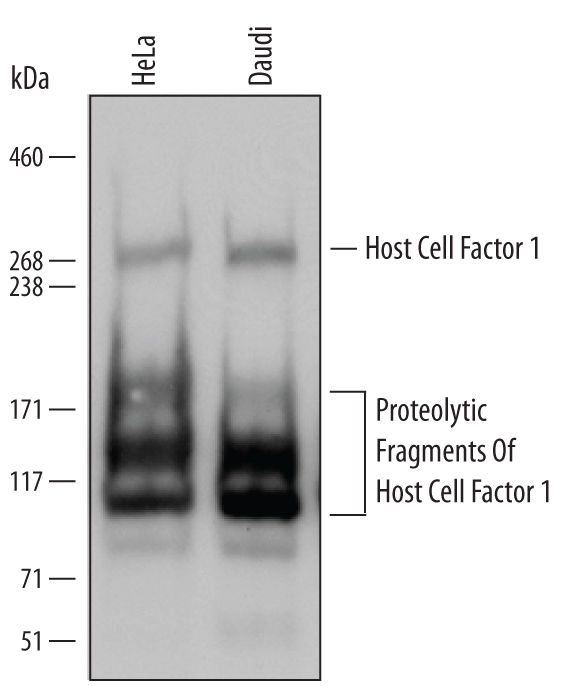 | Detection of Human Host Cell Factor 1/HCFC1 by Western Blot. Western blot shows lysates of HeLa human cervical epithelial carcinoma cell line and Daudi human Burkitt's lymphoma cell line. PVDF Membrane was probed with 1 µg/mL of Goat Anti-Human Host Cell Factor 1/HCFC1 Antigen Affinity-purified Polyclonal Antibody (Catalog # AF6254) followed by HRP-conjugated Anti-Goat IgG Secondary Antibody (Catalog # HAF109). Specific bands were detected for Host Cell Factor 1/HCFC1 at approximately 300 kDa (as indicated) and proteolytic fragments of Host Cell Factor 1/HCFC1 at approximately 100 to 175 kDa (as indicated). This experiment was conducted under reducing conditions and using Immunoblot Buffer Group 1. |
| Immunocytochemistry | |
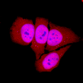 | Host Cell Factor 1/HCFC1 in HeLa Human Cell Line. Host Cell Factor 1/HCFC1 was detected in immersion fixed HeLa human cervical epithelial carcinoma cell line using Goat Anti-Human Host Cell Factor 1/HCFC1 Antigen Affinity-purified Polyclonal Antibody (Catalog # AF6254) at 10 µg/mL for 3 hours at room temperature. Cells were stained using the NorthernLights™ 557-conjugated Anti-Goat IgG Secondary Antibody (red; Catalog # NL001) and counterstained with DAPI (blue). Specific staining was localized to nuclei. View our protocol for Fluorescent ICC Staining of Cells on Coverslips. |
| Immunohistochemistry | |
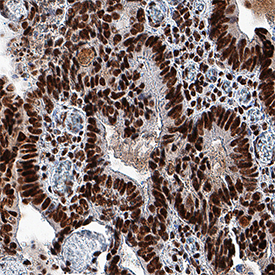 | Host Cell Factor 1/HCFC1 in Human Colon Cancer Tissue. Host Cell Factor 1/HCFC1 was detected in immersion fixed paraffin-embedded sections of human colon cancer tissue using Goat Anti-Human Host Cell Factor 1/HCFC1 Antigen Affinity-purified Polyclonal Antibody (Catalog # AF6254) at 10 µg/mL overnight at 4 °C. Tissue was stained using the Anti-Goat HRP-DAB Cell & Tissue Staining Kit (brown; Catalog # CTS008) and counterstained with hematoxylin (blue). Specific staining was localized to nuclei of epithelial cells. View our protocol for Chromogenic IHC Staining of Paraffin-embedded Tissue Sections. |
追加しました。
Related Product & Information
| Entrez Gene IDs | 3054 (Human); 15161 (Mouse); 363519 (Rat) |
|---|---|
| Background | Host Cell Factor 1/HCFC1 |
| background_content | Background: Host Cell Factor 1/HCFC1 HCF-1 (Host cell factor; also called C1 factor and VCAF) is a blanket name for a group of polypeptides that are generated through the targeted proteolysis of a large 300 kDa transcriptional coactivator precursor. The precursor is widely expressed, and its products are posited to participate in multiple activities, such as mRNA processing, transcriptional coactivation, and cell cycle progression through G0/G1 transition. Human HCF-1 is 2035 amino acids (aa) in length. It is modular in structure, and contains a Kelch-repeat region (aa 32-313), an SP1/GABP basic binding sequence (aa 478-875), an acidic transactivation domain (aa 1530-1735), and a C-terminal NLS-containing Trp/Tyr/Phe-rich region (aa 1760-2035). Situated between the basic and acidic regions is an HCF repeat region (aa 1010-1439) that contains six 26 aa repeats that undergo autocatalytic cleavage. HCF-1 is O-glycosylated, phosphorylated and acetylated. In quiescent cells, the full-length 300 kDa precursor can be found in the cytoplasm, along with a presumed 50 kDa fragment that appears to represent some sequence between aa 300-1000. In activated cells, HCF-1 appears in the nucleus, and undergoes autocatalysis at one or more sites occurs distal to Glu at position 1019, 1081, 1110, 1295, 1323, and 1423. This generates multiple fragments that noncovalently associate with each other and appear as bands in SDS-Page that range from 100-175 kDa in size. This association is quite vigorous and requires aggressive treatment to achieve denaturation. There is one potential isoform that contains an alternative start site at Met100, a second isoform that shows a deletion of aa 1072-1101, a third isoform that exhibits a Leu substitution for aa 428-2035, and a final isoform that shows a deletion of aa 382-450. Over aa 1626-1836, human HCF-1 shares 95% aa identity with mouse HCF-1. |
追加しました。
製品情報は掲載時点のものですが、価格表内の価格については随時最新のものに更新されます。お問い合わせいただくタイミングにより製品情報・価格などは変更されている場合があります。
表示価格に、消費税等は含まれていません。一部価格が予告なく変更される場合がありますので、あらかじめご了承下さい。