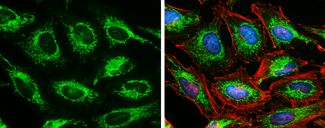
GTX632636 ICC/IF Image
SDHA antibody [GT20710] detects SDHA protein at mitochondria by immunofluorescent analysis.
Sample: HeLa cells were fixed in 4% paraformaldehyde at RT for 15 min.
Green: SDHA protein stained by SDHA antibody [GT20710] (GTX632636) diluted at 1:500.
Red: Phalloidin, a cytoskeleton marker, diluted at 1:100.
Blue: Hoechst 33342 staining.
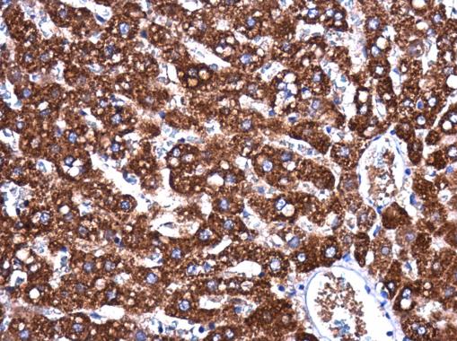
GTX632636 IHC-P Image
SDHA antibody [GT20710] detects SDHA protein at cytoplasm in rat liver by immunohistochemical analysis.
Sample: Paraffin-embedded rat liver.
SDHA antibody [GT20710] (GTX632636) diluted at 1:250.
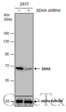
GTX632636 WB Image
Non-transfected (?) and transfected (+) 293T whole cell extracts (30 ug) were separated by 7.5% SDS-PAGE, and the membrane was blotted with SDHA antibody [GT20710] (GTX632636) diluted at 1:1000.
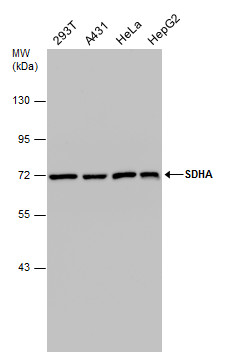
GTX632636 WB Image
Various whole cell extracts (30 ug) were separated by 7.5% SDS-PAGE, and the membrane was blotted with SDHA antibody [GT20710] (GTX632636) diluted at 1:1000.