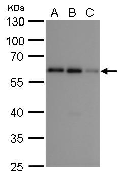
GTX629890 WB Image
SQSTM1 antibody [GT1478] detects SQSTM1 protein by western blot analysis.
A. 30 ug A549 whole cell lysate/extract
B. 30 ug H1299 whole cell lysate/extract
C. 30 ug HCT116 whole cell lysate/extract
10% SDS-PAGE
SQSTM1 antibody [GT1478] (GTX629890) dilution: 1:1000
The HRP-conjugated anti-mouse IgG antibody (GTX213111-01) was used to detect the primary antibody.
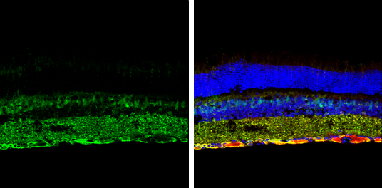
GTX629890 IHC-Fr Image
SQSTM1 antibody [GT1478] detects SQSTM1 protein expression by immunohistochemical analysis.
Sample: Frozen sectioned adult mouse retina.
Green: SQSTM1 protein stained by SQSTM1 antibody [GT1478] (GTX629890) diluted at 1:250.
Red: beta Tubulin 3/ TUJ1, stained by beta Tubulin 3/ TUJ1 antibody [GT11710] (GTX631836) diluted at 1:250.
Blue: Fluoroshield with DAPI (GTX30920).
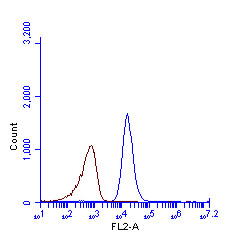
GTX629890 FACS Image
SQSTM1 antibody [GT1478] (GTX629890) detects SQSTM1 protein by flow cytometry analysis.
Sample: HeLa cell fixed in 4% paraformaldehyde at 4oC for 5 min.
Brown: Unlabelled sample was also used as a control.
Blue: SQSTM1 antibody [GT1478] (GTX629890) dilution: 1:100.
Acquisition of >20,000 events were collected using Argon ion laser (488nm) and 525/30 bandpass filter.
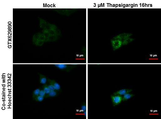
GTX629890 ICC/IF Image
SQSTM1 antibody [GT1478] detects SQSTM1 protein at autophagosome by immunofluorescent analysis.
Samples: HepG2 cells treated with 3uM thapsigargin 16 hrs (rigtht) and mock (left) were fixed in ice-cold MeOH for 10 min, permeabilize with cooled acetone for 1 min .
Green: SQSTM1 protein stained by SQSTM1 antibody [GT1478] (GTX629890) diluted at 1:500.
Blue: Hoechst 33342 staining.
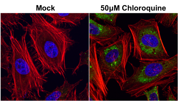
GTX629890 ICC/IF Image
SQSTM1 antibody [GT1478] detects SQSTM1 protein at autophagosome by immunofluorescent analysis.
Samples: HeLa cells mock (left) and treated with 50uM Chloroquine for 24 hr (right) were fixed in 4% paraformaldehyde at RT for 15 min.
Green: SQSTM1 protein stained by SQSTM1 antibody [GT1478] (GTX629890) diluted at 1:1000.
Red: phalloidin, a cytoskeleton marker, stained by phalloidin (invitrogen, A12380) diluted at 1:200.
Blue: Hoechst 33342 staining.
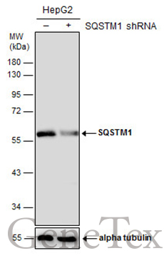
GTX629890 WB Image
Non-transfected (?) and transfected (+) HepG2 whole cell extracts (50 ug) were separated by 10% SDS-PAGE, and the membrane was blotted with SQSTM1 antibody [GT1478] (GTX629890) diluted at 1:500. The HRP-conjugated anti-mouse IgG antibody (GTX213111-01) was used to detect the primary antibody.
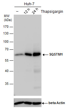
GTX629890 WB Image
SQSTM1 antibody detects SQSTM1 protein by western blot analysis. Un-treated (-) and treated (+, Thapsigargin treatment for 12 hr and 24hr) Huh-7 whole cell extracts (30 ug) were separated by 10% SDS-PAGE, and the membrane was blotted with SQSTM1 antibody (GTX629890) diluted by 1:1000.
The ACTB was used as internal control (GTX110564, 1:50000) shown at the bottom panel.
The HRP-conjugated anti-mouse IgG antibody (GTX213111-01) was used to detect the primary antibody.
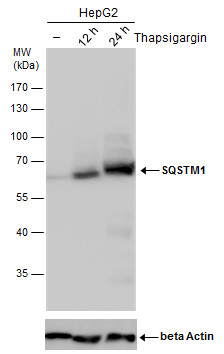
GTX629890 WB Image
SQSTM1 antibody detects SQSTM1 protein by western blot analysis. Un-treated (-) and treated (+, Thapsigargin treatment for 12 hr and 24hr) HepG2 whole cell extracts (30 ug) were separated by 10% SDS-PAGE, and the membrane was blotted with SQSTM1 antibody (GTX629890) diluted by 1:1000.
The ACTB was used as internal control (GTX110564, 1:50000) shown at the bottom panel.
The HRP-conjugated anti-mouse IgG antibody (GTX213111-01) was used to detect the primary antibody.