GTX629744 IHC-Fr Image
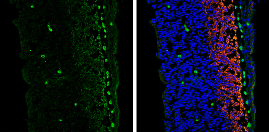
Vimentin antibody [GT812] detects Vimentin protein expression by immunohistochemical analysis.
Sample: Frozen sectioned E13.5 Rat brain.
Green: Vimentin protein stained by Vimentin antibody [GT812] (GTX629744) diluted at 1:250.
Red: beta Tubulin 3/ TUJ1, a mature neuron marker, stained by beta Tubulin 3/ TUJ1 antibody (GTX130245) diluted at 1:250.
Blue: Fluoroshield with DAPI (GTX30920).
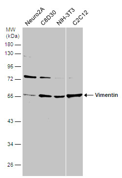
GTX629744 WB Image
Various whole cell extracts (30 ug) were separated by 10% SDS-PAGE, and the membrane was blotted with Vimentin antibody [GT812] (GTX629744) diluted at 1:3000.
GTX629744 WB Image
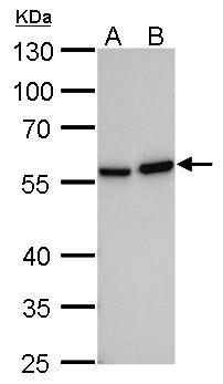
Vimentin antibody [GT812] detects Vimentin protein by Western blot analysis.
A. 30 ug 293T whole cell lysate/extract
B. 30 ug HeLa whole cell lysate/extract
10 % SDS-PAGE
Vimentin antibody [GT812] (GTX629744) dilution: 1:1000
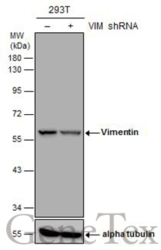
GTX629744 WB Image
Non-transfected (?) and transfected (+) 293T whole cell extracts (30 ug) were separated by 10% SDS-PAGE, and the membrane was blotted with Vimentin antibody [GT812] (GTX629744) diluted at 1:5000.
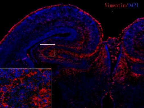
GTX629744 IHC-Fr Image
Vimentin antibody [GT812] detects Vimentin proteins in embryonic mouse brain by immunohistochemical analysis.
Sample: Frozen section of embryonic mouse brain (mE18.5).
Red: Vimentin antibody [GT812] (GTX629744) diluted at 1:250.
Blue: DAPI.
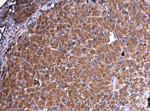
GTX629744 IHC-P Image
Vimentin antibody [GT812] detects Vimentin protein at cytoplasm in rat ovary by immunohistochemical analysis.
Sample: Paraffin-embedded rat ovary.
Vimentin antibody [GT812] (GTX629744) diluted at 1:250.
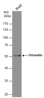
GTX629744 WB Image
Whole cell extract (30 ug) was separated by 10% SDS-PAGE, and the membrane was blotted with Vimentin antibody [GT812] (GTX629744) diluted at 1:75000.
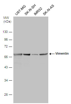
GTX629744 WB Image
Various whole cell extracts (30 ug) were separated by 10% SDS-PAGE, and the membrane was blotted with Vimentin antibody [GT812] (GTX629744) diluted at 1:5000.