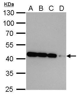
GTX629630 WB Image
Beta Actin antibody [GT5512] detects beta Actin protein by western blot analysis.
A. 20 ug 293T whole cell lysate/extract
B. 10 ug 293T whole cell lysate/extract
C. 5 ug 293T whole cell lysate/extract
D. 1 ug 293T whole cell lysate/extract
10% SDS-PAGE
Beta Actin antibody [GT5512] (GTX629630) dilution: 1:10000
The HRP-conjugated anti-mouse IgG antibody (GTX213111-01) was used to detect the primary antibody.
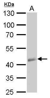
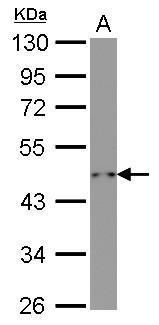
GTX629630 WB Image
beta Actin antibody [GT5512] detects beta Actin protein by western blot analysis.
A. 30 ug 30 hpf zebrafish lysate/extract
10% SDS-PAGE
beta Actin antibody [GT5512] (GTX629630) dilution: 1:1000
The HRP-conjugated anti-mouse IgG antibody (GTX213111-01) was used to detect the primary antibody.
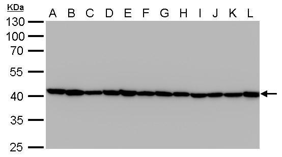
GTX629630 WB Image
Beta Actin antibody [GT5512] detects beta Actin protein by western blot analysis.
A. 30 ug Jurkat whole cell lysate/extract
B. 30 ug Raji whole cell lysate/extract
C. 30 ug 293T whole cell lysate/extract
D. 30 ug A431 whole cell lysate/extract
E. 30 ug HeLa whole cell lysate/extract
F. 30 ug HepG2 whole cell lysate/extract
G. 30 ug H1299 whole cell lysate/extract
H. 30 ug HCT116 whole cell lysate/extract
I. 30 ug MCF-7 whole cell lysate/extract
J. 30 ug NT2D1 whole cell lysate/extract
K. 30 ug PC-3 whole cell lysate/extract
L. 30 ug U87-MG whole cell lysate/extract
10% SDS-PAGE
Beta Actin antibody [GT5512] (GTX629630) dilution: 1:20000
The HRP-conjugated anti-mouse IgG antibody (GTX213111-01) was used to detect the primary antibody.
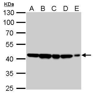
GTX629630 WB Image
Beta Actin antibody [GT5512] detects beta Actin protein by western blot analysis.
A. 30 ug 293T whole cell lysate/extract
B. 30 ug NIH-3T3 whole cell lysate/extract
C. 30 ug mouse brain lysate/extract
D. 30 ug PC-12 whole cell lysate/extract
E. 30 ug rat brain lysate/extract
10% SDS-PAGE
Beta Actin antibody [GT5512] (GTX629630) dilution: 1:10000
The HRP-conjugated anti-mouse IgG antibody (GTX213111-01) was used to detect the primary antibody.
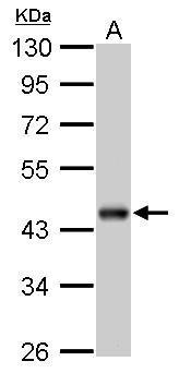
GTX629630 WB Image
beta Actin antibody [GT5512] detects beta Actin protein by western blot analysis.
A. 10 ug yeast lysate/extract
10% SDS-PAGE
beta Actin antibody [GT5512] (GTX629630) dilution: 1:1000
The HRP-conjugated anti-mouse IgG antibody (GTX213111-01) was used to detect the primary antibody.
GTX629630 WB Image
Beta Actin antibody [GT5512] detects beta Actin protein by western blot analysis.
A. 30 ug drosophila lysate/extract
10% SDS-PAGE
Beta Actin antibody [GT5512] (GTX629630) dilution: 1:1000
The HRP-conjugated anti-mouse IgG antibody (GTX213111-01) was used to detect the primary antibody.
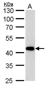
GTX629630 WB Image
beta Actin antibody [GT5512] detects beta Actin protein by western blot analysis.
A. 30 ug rabbit blood lysate/extract
10% SDS-PAGE
beta Actin antibody [GT5512] (GTX629630) dilution: 1:10000
The HRP-conjugated anti-mouse IgG antibody (GTX213111-01) was used to detect the primary antibody.
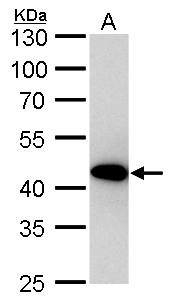
GTX629630 WB Image
beta Actin antibody [GT5512] detects beta Actin protein by western blot analysis.
A. 30 ug goat blood lysate/extract
10% SDS-PAGE
beta Actin antibody [GT5512] (GTX629630) dilution: 1:10000
The HRP-conjugated anti-mouse IgG antibody (GTX213111-01) was used to detect the primary antibody.
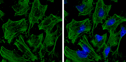
GTX629630 ICC/IF Image
beta Actin antibody [GT5512] detects beta Actin protein at cytoskeleton by immunofluorescent analysis.
Sample: HeLa cells were fixed in 0.5% Triton X-100 for 1 min, then ice-cold methanol for 5 min.
Green: beta Actin protein stained by beta Actin antibody [GT5512] (GTX629630) diluted at 1:500.
Blue: Hoechst 33342 staining.
GTX629630 WB Image
beta Actin antibody[GT5512] detects beta Actin protein by western blot analysis.
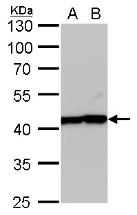
A. 30 ug MCF-7 whole cell lysate/extract
B. 30 ug MDA-MB-231 whole cell lysate/extract
10% SDS-PAGE
beta Actin antibody[GT5512] (GTX629630) dilution: 1:20000
The HRP-conjugated anti-mouse IgG antibody (GTX213111-01) was used to detect the primary antibody.
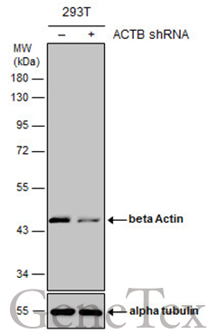
GTX629630 WB Image
Non-transfected (?) and transfected (+) 293T whole cell extracts (10 ug) were separated by 10% SDS-PAGE, and the membrane was blotted with beta Actin antibody [GT5512] (GTX629630) diluted at 1:20000. The HRP-conjugated anti-mouse IgG antibody (GTX213111-01) was used to detect the primary antibody.
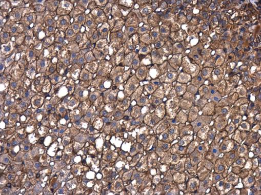
GTX629630 IHC-P Image
beta Actin antibody [GT5512] detects beta Actin protein at cell membrane and cytoplasm in rat adrenal gland by immunohistochemical analysis.
Sample: Paraffin-embedded rat adrenal gland.
beta Actin antibody [GT5512] (GTX629630) diluted at 1:250.