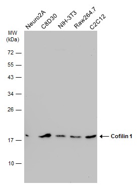
GTX628804 WB Image
Various whole cell extracts (30 ug) were separated by 12% SDS-PAGE, and the membrane was blotted with Cofilin 1 antibody [GT567] (GTX628804) diluted at 1:1000. The HRP-conjugated anti-mouse IgG antibody (GTX213111-01) was used to detect the primary antibody.
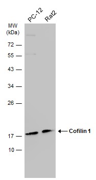
GTX628804 WB Image
Various whole cell extracts (30 ug) were separated by 12% SDS-PAGE, and the membrane was blotted with Cofilin 1 antibody [GT567] (GTX628804) diluted at 1:1000. The HRP-conjugated anti-mouse IgG antibody (GTX213111-01) was used to detect the primary antibody.
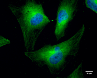
GTX628804 ICC/IF Image
Cofilin 1 antibody [GT567] detects Cofilin 1 protein at cytoplasm by immunofluorescent analysis.
Sample: HeLa cells were fixed in 4% paraformaldehyde at RT for 10 min.
Green: Cofilin 1 protein stained by Cofilin 1 antibody [GT567] (GTX628804) diluted at 1:300.
Blue: Hoechst 33342 staining.
Scale bar = 10 um.
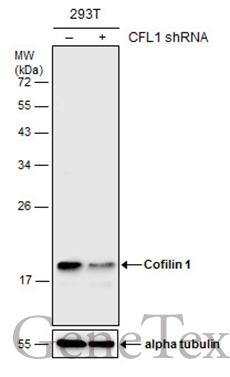
GTX628804 WB Image
Non-transfected (?) and transfected (+) 293T whole cell extracts (30 ug) were separated by 12% SDS-PAGE, and the membrane was blotted with Cofilin 1 antibody [GT567] (GTX628804) diluted at 1:500. The HRP-conjugated anti-mouse IgG antibody (GTX213111-01) was used to detect the primary antibody.

GTX628804 IHC-P Image
Cofilin 1 antibody [GT567] detects CFL1 protein at cytosol on HBL435 xenograft by immunohistochemical analysis.
Sample: Paraffin-embedded HBL435 xenograft.
Cofilin 1 antibody [GT567] (GTX628804) dilution: 1:200.
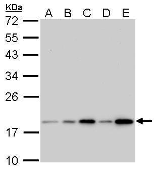
GTX628804 WB Image
Cofilin 1 antibody [GT567] detects CFL1 protein by western blot analysis.
A. 30 ug 293T whole cell lysate/extract
B. 30 ug A431 whole cell lysate/extract
C. 30 ug HeLa whole cell lysate/extract
D. 30 ug HepG2 whole cell lysate/extract
E. 30 ug A375 whole cell lysate/extract
12% SDS-PAGE
Cofilin 1 antibody [GT567] (GTX628804) dilution: 1:1000
The HRP-conjugated anti-mouse IgG antibody (GTX213111-01) was used to detect the primary antibody.