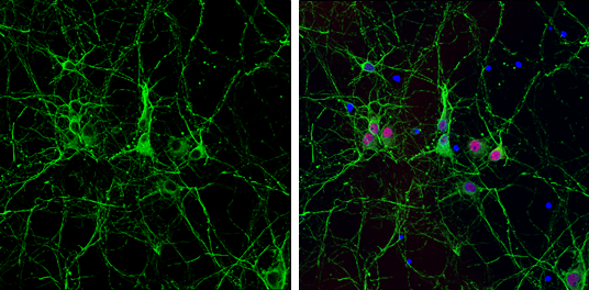
GTX628802 ICC/IF Image
alpha Tubulin antibody [GT114] detects alpha Tubulin protein expression by immunofluorescent analysis.
Sample: Cultured rat E18 primary cortical neuron, DIV 8. Cells were fixed in 4% paraformaldehyde at RT for 15 min.
Green: alpha Tubulin protein stained by alpha Tubulin antibody [GT114] (GTX628802) diluted at 1:250.
Red: NeuN, stained by NeuN antibody (GTX132974) diluted at 1:250.
Blue: Fluoroshield with DAPI (GTX30920).
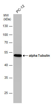
GTX628802 WB Image
Whole cell extract (30 ug) was separated by 10% SDS-PAGE, and the membrane was blotted with alpha Tubulin antibody [GT114] (GTX628802) diluted at 1:10000. The HRP-conjugated anti-mouse IgG antibody (GTX213111-01) was used to detect the primary antibody.
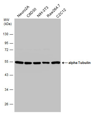
GTX628802 WB Image
Various whole cell extracts (30 ug) were separated by 10% SDS-PAGE, and the membrane was blotted with alpha Tubulin antibody [GT114] (GTX628802) diluted at 1:10000. The HRP-conjugated anti-mouse IgG antibody (GTX213111-01) was used to detect the primary antibody.
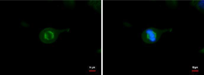
GTX628802 ICC/IF Image
TUBA1B antibody detects TUBA1B protein at Cytoskeleton by immunofluorescent analysis.
Sample: HeLa cells were fixed in -20Åé 100% methanol for 5 min.
Green: TUBA1B protein stained by TUBA1B antibody (GTX628802) diluted at 1:500.
Blue: Hoechst 33342 staining.
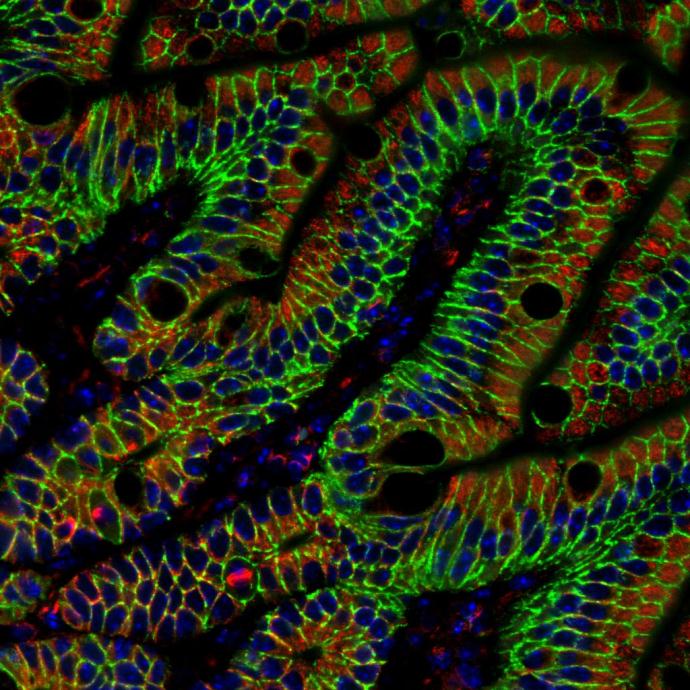
GTX628802 IHC-P Image
alpha Tubulin antibody [GT114] detects alpha Tubulin protein at cytoplasm in mouse colon by immunohistochemical analysis.
Sample: Paraffin-embedded mouse colon.
Red: alpha Tubulin antibody [GT114] (GTX628802) diluted at 1:500.
Green: beta Catenin antibody [N1N2-2] (GTX101435) diluted at 1:500.
Blue: Hoechst 33342 staining.
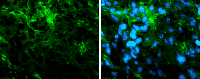
GTX628802 IHC-Fr Image
Immunofluorescence photomicrographs of frozen sections of mouse brain.
Green: alpha Tubulin antibody [GT114] (GTX628802) diluted at 1:200. The signal was developed using goat anti-rmouse IgG antibody (Dylight488) (GTX213111-04).
Blue: Nuclear staining with Hoechst 33342.
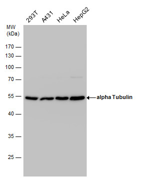
GTX628802 WB Image
alpha Tubulin antibody detects alpha Tubulin protein by western blot analysis. Various whole cell extracts (30 ug) were separated by 10% SDS-PAGE, and the membrane was blotted with alpha Tubulin antibody (GTX628802) diluted at a dilution of 1:10000. The HRP-conjugated anti-mouse IgG antibody (GTX213111-01) was used to detect the primary antibody.
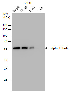
GTX628802 WB Image
alpha Tubulin antibody detects alpha Tubulin protein by western blot analysis. Varied amount of 293T Whole cell extracts was separated by 10% SDS-PAGE, and the membrane was blotted with alpha Tubulin antibody (GTX628802) at a dilution of 1:10000. The HRP-conjugated anti-mouse IgG antibody (GTX213111-01) was used to detect the primary antibody.

GTX628802 WB Image
alpha Tubulin antibody [GT114] detects TUBA1B protein by western blot analysis.
A. 30 ug 293T whole cell lysate/extract
B. 30 ug NIH-3T3 whole cell lysate/extract
C. 30 ug mouse brain lysate/extract
D. 30 ug PC-12 whole cell lysate/extract
E. 30 ug rat brain lysate/extract
F. 30 ug drosophila lysae/extract
10% SDS-PAGE
alpha Tubulin antibody [GT114] (GTX628802) dilution: 1:5000
The HRP-conjugated anti-mouse IgG antibody (GTX213111-01) was used to detect the primary antibody.
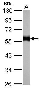
GTX628802 WB Image
alpha Tubulin antibody [GT114] detects TUBA1B protein by western blot analysis.
A. 30 ug zebrafish eye lysate/extract
10% SDS-PAGE
alpha Tubulin antibody [GT114] (GTX628802) dilution: 1:1000
The HRP-conjugated anti-mouse IgG antibody (GTX213111-01) was used to detect the primary antibody.
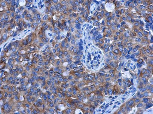
GTX628802 IHC-P Image
alpha Tubulin antibody [GT114] detects alpha Tubulin protein at cytoplasm in human lung cancer by immunohistochemical analysis.
Sample: Paraffin-embedded human lung cancer.
alpha Tubulin antibody [GT114] (GTX628802) diluted at 1:500.
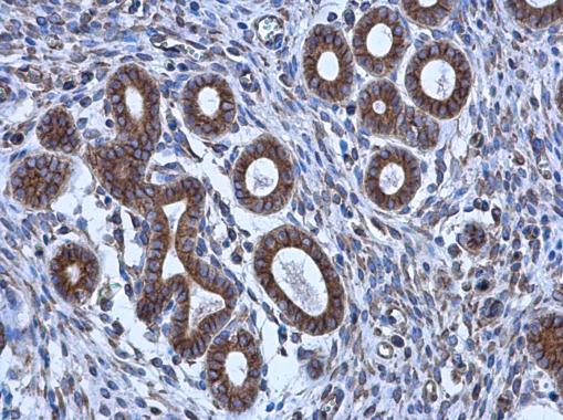
GTX628802 IHC-P Image
alpha Tubulin antibody [GT114] detects alpha Tubulin protein at cytoplasm in human cervix by immunohistochemical analysis.
Sample: Paraffin-embedded human cervix.
alpha Tubulin antibody [GT114] (GTX628802) diluted at 1:500.

GTX628802 ICC/IF Image
alpha Tubulin antibody [GT114] detects alpha Tubulin protein at cytoskeleton by immunofluorescent analysis.
Sample: HeLa cells were fixed in 4% paraformaldehyde at RT for 15 min.
Green: alpha Tubulin protein stained by alpha Tubulin antibody [GT114] (GTX628802) diluted at 1:1000.
Blue: Hoechst 33342 staining.
Scale bar = 10 um.