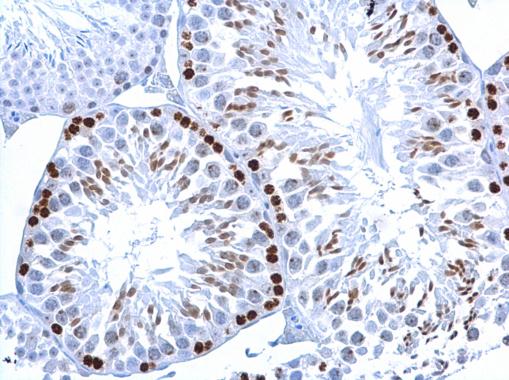
GTX628789 IHC-P Image
Histone H2A.XS139ph (phospho Ser139) antibody [GT2311] detects Histone H2A.XS139ph (phospho Ser139) protein at nucleus on mouse testis by immunohistochemical analysis.
Sample: Paraffin-embedded mouse testis.
Histone H2A.XS139ph (phospho Ser139) antibody [GT2311] (GTX628789) dilution: 1:500.
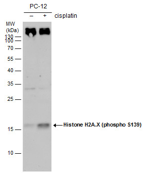
GTX628789 WB Image
Histone H2A.X (phospho S139) antibody [GT2311] detects Histone H2A.X (phospho S139) [GT2311] protein by western blot analysis. Un-treated (-) and treated (+, 30 uM Cisplatin treatment for 24 hrs) PC-12 whole cell extracts (30 ug) were separated by 15% SDS-PAGE, and the membrane was blotted with Histone H2A.X (phospho S139) antibody [GT2311] (GTX628789) diluted by 1:500. The HRP-conjugated anti-mouse IgG antibody (GTX213111-01) was used to detect the primary antibody.
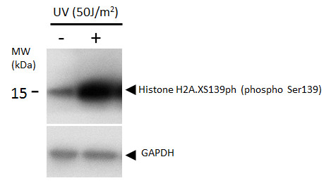
GTX628789 WB Image
Histone H2A.XS139ph (phospho Ser139) antibody detects Histone H2A.XS139ph (phospho Ser139) protein by western blot analysis. Un-treated (-) and treated (+, 50 J/m2 UV treatment) U2OS whole cell extracts (16 ug) were separated by 12%-15% SDS-PAGE, and the membrane was blotted with Histone H2A.XS139ph (phospho Ser139) antibody (GTX628789) diluted at 1:1000. The HRP-conjugated anti-mouse IgG antibody (GTX213111-01) was used to detect the primary antibody.
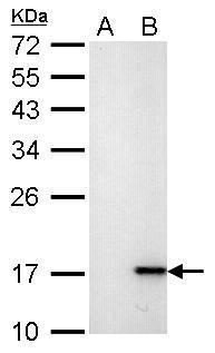
GTX628789 WB Image
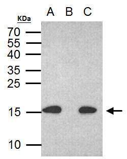
Histone H2A.X (phospho S139) antibody [GT2311] detects H2AFX protein by western blot analysis.
A. 30 ug HCT116 whole cell lysate/extract (untreated)
B. 30 ug HCT116 whole cell lysate/extract (30 uM cisplatin treatment for 24hr)
12% SDS-PAGE
Histone H2A.X (phospho S139) antibody [GT2311] (GTX628789) dilution: 1:1000
The HRP-conjugated anti-mouse IgG antibody (GTX213111-01) was used to detect the primary antibody.
GTX628789 ICC/IF Image
Histone H2A.X (phospho Ser139) antibody detects H2AFX protein at nuclear by confocal immunofluorescent analysis.
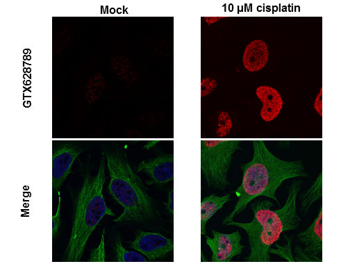
Sample: 10uM Cisplatin treated (right) or untreated (left) HeLa cells were fixed in 4% paraformaldehyde for 15 min.
Red: H2A.X protein stained by Histone H2A.X (phospho Ser139) antibody (GTX628789) diluted at 1:500.
Green: alpha Tubulin antibody (GTX102078) diluted at 1:1000.
Blue: Hoechst 33342 staining. [Images captured by Olympus FV1000 Confocal Laser Scanning Microscope]
GTX628789 ICC/IF Image
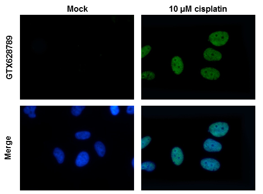
Histone H2A.X antibody detects H2AFX protein at nuclear by immunofluorescent analysis. Sample: 10uM Cisplatin treated (right) or untreated (left) HeLa cells were fixed in 4% paraformaldehyde for 15 min. Green: H2AFX protein stained by Histone H2A.Xantibody (GTX628789) diluted at 1:500. Blue: Hoechst 33342 staining.
GTX628789 IP Image
Histone H2A.X (phospho S139) antibody immunoprecipitates histone H2A.X (phospho S139) protein in IP experiments. IP Sample: 500 ug HCT116 with CPT 30 uM treatment 24 hr whole cell lysate/extract A. 30 ug HCT116 whole with CPT 30 uM treatment cell lysate/extract B. Control with 2 ug of preimmune mouse IgG C. Immunoprecipitation of histone H2A.X (phospho S139) protein by 2 ug histone H2A.X (phospho S139) antibody (GTX628789) 15% SDS-PAGE The immunoprecipitated histone H2A.X (phospho S139) protein was detected by Human histone H2A.X (phospho S139) antibody (GTX628789) diluted at 1:1000. EasyBlot anti-mouse IgG (GTX221667-01) was used as a secondary reagent.
GTX628789 WB Image
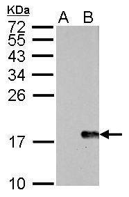
Histone H2A.X (phospho S139) antibody [GT2311] detects H2AFX protein by western blot analysis.
A. 30 ug NIH-3T3 whole cell lysate/extract (untreated)
B. 30 ug NIH-3T3 whole cell lysate/extract (30uM cisplatin treatment for 24hr)
15% SDS-PAGE
Histone H2A.X (phospho S139) antibody [GT2311] (GTX628789) dilution: 1:1000
The HRP-conjugated anti-mouse IgG antibody (GTX213111-01) was used to detect the primary antibody.