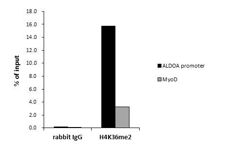
GTX60372 ChIP Image
ChIP was performed with HeLa nuclear extract and either 15 ul of H4K8ac antibody or 5 ug of control rabbit IgG. The precipitated DNA was detected by QPCR with primer set targeting to ALDOA promoter or MyoD.
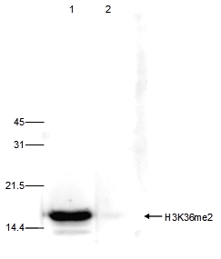
GTX60372 WB Image
WB analysis of histone extracts (15 ug) from HeLa cells using H3K36me2 antibody diluted 1:1,000 in TBS-Tween containing 5% skimmed milk (lane 1). Lane 2 shows the same analysis after incubation of the antibody with 5 nmol blocking peptide for 1 hour at room temperature.
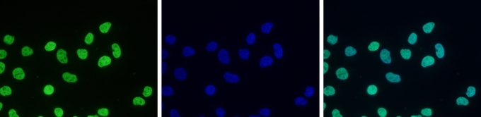
GTX60372 ICC/IF Image
ICC/IF analysis of HeLa cells using H3K36me2 antibody and DAPI. Cells were fixed with 4% formaldehyde for 10Åf and blocked with PBS/TX-100 containing 5% normal goat serum and 1% BSA. The cells were immunofluorescently labelled with the H3K36me2 antibody at a dilution of 1:500 (green). The nuclei were stained with DAPI (blue).

GTX60372 Dot Image
Dot blot analysis of peptides containing modified and unmodified sequences of histone H3 using H3K36me2 antibody at a dilution of 1:100,000. One hundred to 0.2 pmol of the peptide containing the respective histone modification were spotted on a membrane for analysis.
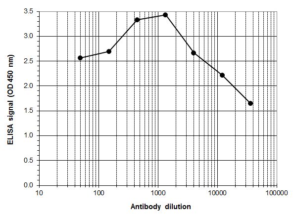
GTX60372 ELISA Image
ELISA was performed using a serial dilution of Histone H3K36me2 (di-Methyl Lys36) antibody in antigen coated wells. By plotting the absorbance against the antibody dilution, the titer of the antibody was estimated to be 1:31,000.