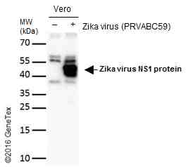
GTX133307 WB Image
Non-infected (?) and infected (+) vero cells (15 ug) were separated by gradient gel, and the membrane was blotted with Zika virus NS1 protein antibody (GTX133307) diluted at 1:2000. The HRP-conjugated anti-rabbit IgG antibody (GTX213110-01) was used to detect the primary antibody.
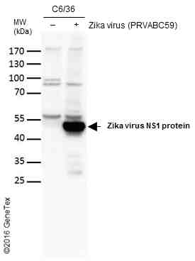
GTX133307 WB Image
Non-infected (?) and infected (+) C6/36 cells (20 ug) were separated by gradient gel, and the membrane was blotted with Zika virus NS1 protein antibody (GTX133307) diluted at 1:4000. The HRP-conjugated anti-rabbit IgG antibody (GTX213110-01) was used to detect the primary antibody.
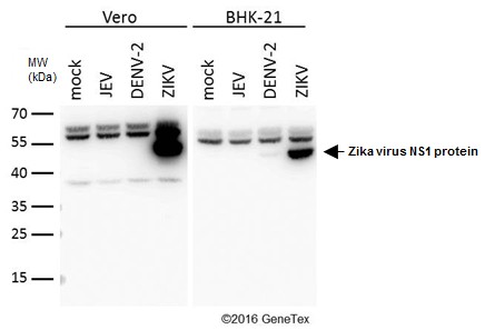
GTX133307 WB Image
Mock and infected Vero and BHK-21 whole cell extracts (20 ug) were separated by gradient gel, and the membrane was blotted with Zika virus NS1 protein antibody (GTX133307) diluted at 1:4000.
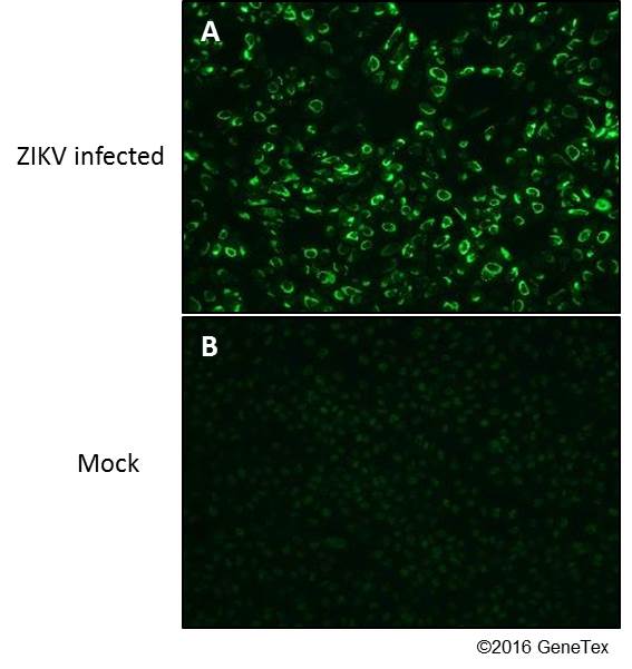
GTX133307 ICC/IF Image
Immunofluorescent analysis of Zika Virus-PRVABC59 infected (A) and non-infected (B) vero cells using Zika virus NS1 protein antibody (GTX133307).
Green: Zika virus NS1 protein antibody (GTX133307) diluted at 1:4000.
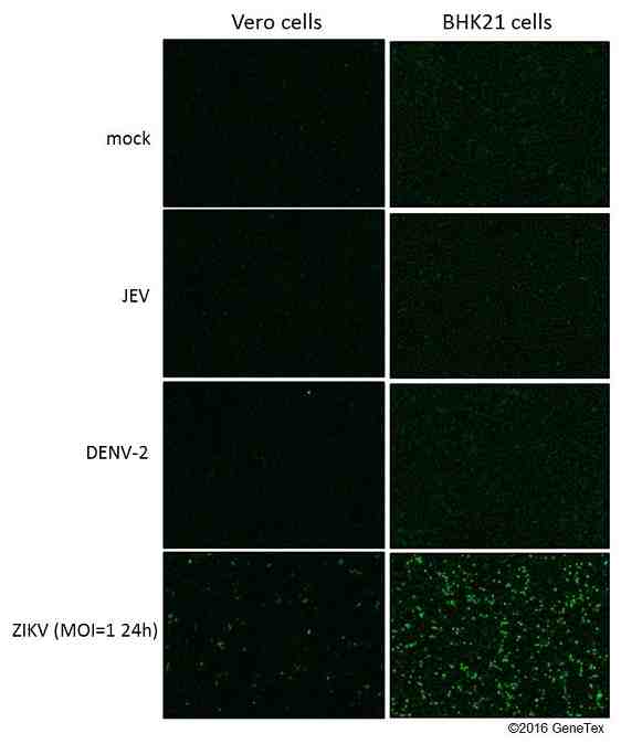
GTX133307 ICC/IF Image
Immunofluorescent analysis of non-infected and infected vero or BHK-21 cells using Zika virus NS1 protein antibody (GTX133307).
Green: Zika virus NS1 protein antibody (GTX133307) diluted at 1:4000.
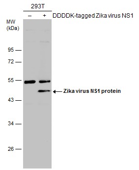
GTX133307 WB Image
Non-transfected (?) and transfected (+) 293T whole cell extracts (30 ug) were separated by 10% SDS-PAGE, and the membrane was blotted with Zika virus NS1 protein antibody (GTX133307) diluted at 1:200. The HRP-conjugated anti-rabbit IgG antibody (GTX213110-01) was used to detect the primary antibody.