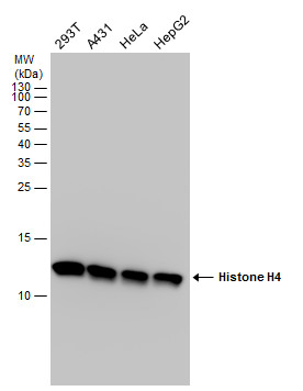
GTX129560 WB Image
Histone H4 antibody detects Histone H4 protein by western blot analysis. Various whole cell extracts (30 ug) were separated by 15% SDS-PAGE, and the membrane was blotted with Histone H4 antibody (GTX129560) diluted at a dilution of 1:5000.
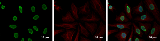
GTX129560 ICC/IF Image
Histone H4 antibody detects Histone H4 protein at nucleus by immunofluorescent analysis.
Sample: HeLa cells were fixed in 4% paraformaldehyde at RT for 15 min.
Green: Histone H4 protein stained by Histone H4 antibody (GTX129560) diluted at 1:5000.
Red: alpha Tubulin, a cytoskeleton marker, stained by alpha Tubulin antibody [GT114] (GTX628802) diluted at 1:2000.
Blue: Hoechst 33342 staining.

GTX129560 WB Image
Various tissue extracts (30 ug) were separated by 15% SDS-PAGE, and the membrane was blotted with Histone H4 antibody (GTX129560) diluted at 1:1000.
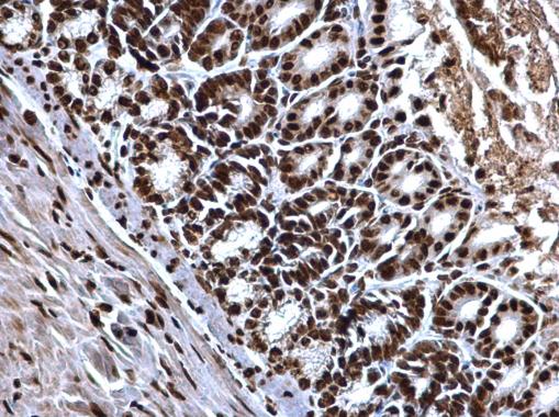
GTX129560 IHC-P Image
Histone H4 antibody detects Histone H4 protein at nucleus on mouse duodenum by immunohistochemical analysis.
Sample: Paraffin-embedded mouse duodenum.
Histone H4 antibody (GTX129560) dilution: 1:500.
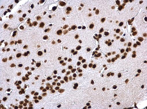
GTX129560 IHC-P Image
Histone H4 antibody detects Histone H4 protein at nucleus on mouse fore brain by immunohistochemical analysis.
Sample: Paraffin-embedded mouse fore brain.
Histone H4 antibody (GTX129560) dilution: 1:500.
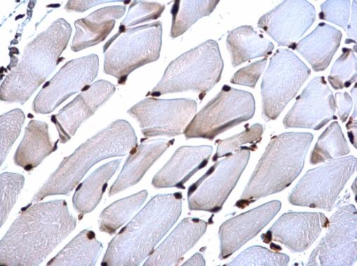
GTX129560 IHC-P Image
Histone H4 antibody detects Histone H4 protein at nucleus on mouse muscle by immunohistochemical analysis.
Sample: Paraffin-embedded mouse muscle.
Histone H4 antibody (GTX129560) dilution: 1:500.
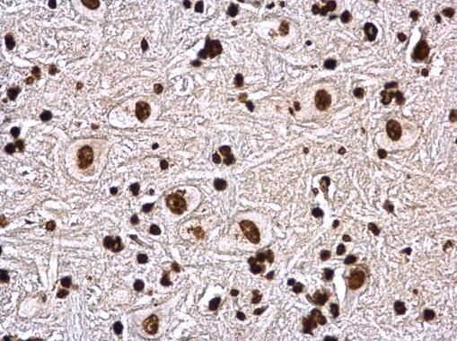
GTX129560 IHC-P Image
Histone H4 antibody detects Histone H4 protein at nucleus on rat brain stem by immunohistochemical analysis.
Sample: Paraffin-embedded rat brain stem.
Histone H4 antibody (GTX129560) dilution: 1:500.