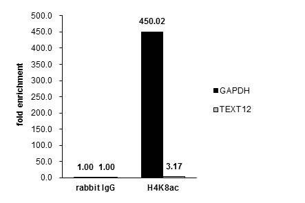
GTX128957 ChIP assay Image
Cross-linked ChIP was performed with HeLa chromatin extract treated with Trichostatin A (0.4 uM for 18 h) and 5 ug of either control rabbit IgG or anti-Histone H4K8ac (acetyl Lys8) antibody. The precipitated DNA was detected by PCR with primer set targeting to GAPDH or TEXT12.

GTX128957 IP Image
Histone H4 (acetyl K9) antibody immunoprecipitates Histone H4 (acetyl K9) protein in IP experiments. IP Sample: HeLa whole cell lysate/extract A : 30 ug whole cell lysate/extract of Histone H4 (acetyl K9) protein expressing HeLa cells (0.4uM TSA treatment for 18hr) B : Control with 2.5 ug of pre-immune rabbit IgG C : Immunoprecipitation of Histone H4 (acetyl K9) protein by 2.5 ug of Histone H4 (acetyl K9) antibody (GTX128957). 12% SDS-PAGE The immunoprecipitated Histone H4 (acetyl K9) protein was detected by Histone H4 (acetyl K9) antibody (GTX128957) diluted at 1 : 5000. EasyBlot anti-rabbit IgG (HRP) (GTX221666-01) was used as a secondary reagent.
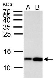
GTX128957 WB Image
Histone H4 (acetyl Lys8) antibody detects Histone H4 (acetyl Lys8) protein by Western blot analysis.
A. 30 ug HeLa whole cell lysate/extract (0.012% DMSO treatment for 18 hr)
B. 30 ug HeLa whole cell lysate/extract (0.4 uM Trichostatin A treatment for 18 hr)
15 % SDS-PAGE
Histone H4 (acetyl Lys8) antibody (GTX128957) dilution: 1:5000

GTX128957 IHC-P Image
Histone H4K8ac (acetyl Lys8) antibody detects Histone H4K8ac (acetyl Lys8) protein at nucleus on mouse duodenum by immunohistochemical analysis.
Sample: Paraffin-embedded mouse duodenum.
Histone H4K8ac (acetyl Lys8) antibody (GTX128957) dilution: 1:500.
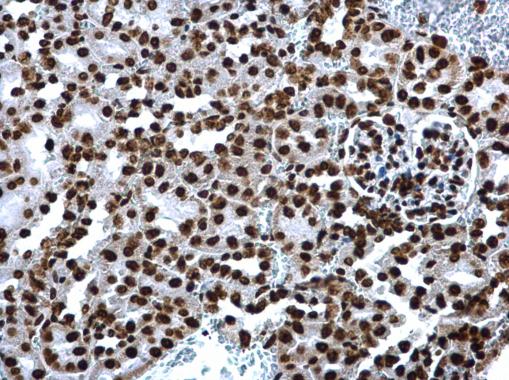
GTX128957 IHC-P Image
Histone H4K8ac (acetyl Lys8) antibody detects Histone H4K8ac (acetyl Lys8) protein at nucleus on mouse kidney by immunohistochemical analysis.
Sample: Paraffin-embedded mouse kidney.
Histone H4K8ac (acetyl Lys8) antibody (GTX128957) dilution: 1:500.
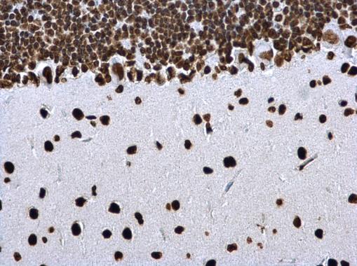
GTX128957 IHC-P Image
Histone H4K8ac (acetyl Lys8) antibody detects Histone H4K8ac (acetyl Lys8) protein at nucleus on rat hind brain by immunohistochemical analysis.
Sample: Paraffin-embedded rat hind brain.
Histone H4K8ac (acetyl Lys8) antibody (GTX128957) dilution: 1:500.

GTX128957 Dot Image
Dotblot analysis of anti-Histone H4 (acetyl Lys9) antibody with peptide samples.
Varied amount of peptide samples were spotted onto positively charged nylon membrane and blotted with Histone H4 (acetyl Lys9) antibody (GTX128957) 1:5000 dilution.
A: Peptide samples of H4K8 un
B: Peptide samples of H4K8ac
C: Peptide samples of H4K16un
D: Peptide samples of H4K16ac
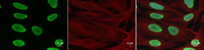
GTX128957 ICC/IF Image
Histone H4 (acetyl Lys8) antibody detects Histone H4 (acetyl Lys8) protein at nucleus by immunofluorescent analysis.
Sample: HeLa cells were fixed in 4% paraformaldehyde at RT for 15 min.
Green: Histone H4 (acetyl Lys8) protein stained by Histone H4 (acetyl Lys8) antibody (GTX128957) diluted at 1:500.
Red: alpha Tubulin antibody [GT114], a cytoskeleton marker, stained by GTX628802 diluted at 1:500.
Blue: Hoechst 33342 staining.