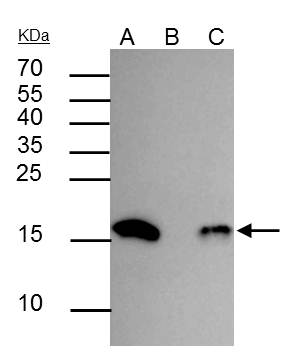
GTX128943 IP Image
Histone H3 (acetyl Lys18) antibody immunoprecipitates Histone H3 (acetyl Lys18) protein in IP experiments. IP Sample: HeLa whole cell lysate/extract A : 30 ug whole cell lysate/extract of Histone H3 (acetyl Lys18) protein expressing HeLa cells (0.4uM TSA treatment for 18hr) B : Control with 2.5 ug of pre-immune rabbit IgG C : Immunoprecipitation of Histone H3 (acetyl Lys18) protein by 2.5 ug of Histone H3 (acetyl Lys18) antibody (GTX128943) 12% SDS-PAGE The immunoprecipitated Histone H3 protein (acetyl Lys18) was detected by Histone H3 (acetyl Lys18) antibody (GTX128943) diluted at 1 : 5000. EasyBlot anti-rabbit IgG (HRP) (GTX221666-01) was used as a secondary reagent.
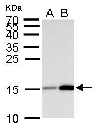
GTX128943 WB Image
Histone H3 (acetyl Lys18) antibody detects Histone H3 (acetyl Lys18) protein by Western blot analysis.
A. 30 ug HeLa whole cell lysate/extract (0.012% DMSO treatment for 18 hr)
B. 30 ug HeLa whole cell lysate/extract (0.4 uM Trichostatin A treatment for 18 hr)
15 % SDS-PAGE
Histone H3 (acetyl Lys18) antibody (GTX128943) dilution: 1:5000
GTX128943 IHC-P Image
Histone H3K18ac (acetyl Lys18) antibody detects Histone H3K18ac (acetyl Lys18) protein at nucleus on mouse duodenum by immunohistochemical analysis.
Sample: Paraffin-embedded mouse duodenum.
Histone H3K18ac (acetyl Lys18) antibody (GTX128943) dilution: 1:500.
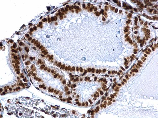
GTX128943 IHC-P Image
Histone H3K18ac (acetyl Lys18) antibody detects Histone H3K18ac (acetyl Lys18) protein at nucleus on mouse prostate by immunohistochemical analysis.
Sample: Paraffin-embedded mouse prostate.
Histone H3K18ac (acetyl Lys18) antibody (GTX128943) dilution: 1:500.
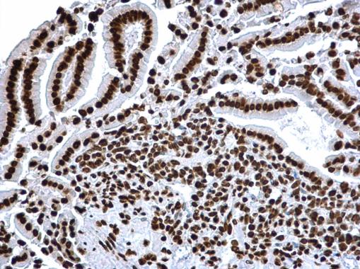
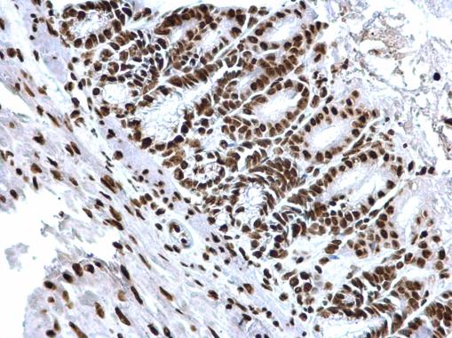
GTX128943 IHC-P Image
Histone H3K18ac (acetyl Lys18) antibody detects Histone H3K18ac (acetyl Lys18) protein at nucleus on mouse intestine by immunohistochemical analysis.
Sample: Paraffin-embedded mouse intestine.
Histone H3K18ac (acetyl Lys18) antibody (GTX128943) dilution: 1:500.
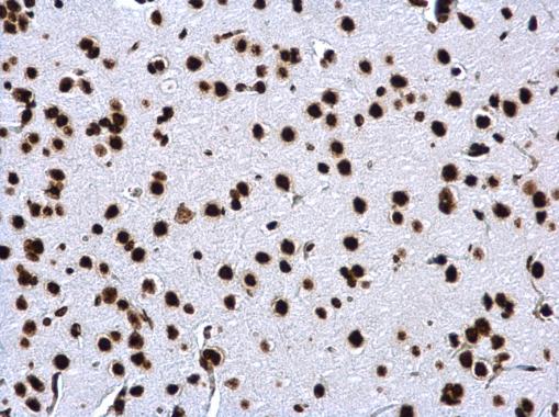
GTX128943 IHC-P Image
Histone H3K18ac (acetyl Lys18) antibody detects Histone H3K18ac (acetyl Lys18) protein at nucleus on rat fore brain by immunohistochemical analysis.
Sample: Paraffin-embedded rat fore brain.
Histone H3K18ac (acetyl Lys18) antibody (GTX128943) dilution: 1:500.

GTX128943 Dot Image
Dotblot analysis of anti-Histone H3 (acetyl Lys18) antibody with peptide samples.
Varied amount of peptide samples were spotted onto positively charged nylon membrane and blotted with Histone H3 (acetyl Lys18) antibody (GTX128943) 1:5000 dilution.
A: Peptide samples of H3K18 un
B: Peptide samples of H3K18ac
C: Peptide samples of H3K9un
D: Peptide samples of H3K9ac
E: Peptide samples of H3K14un
F: Peptide samples of H3K14ac
G: Peptide samples of H3K27un
H: Peptide samples of H3K27ac
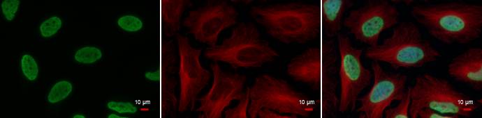
GTX128943 ICC/IF Image
Histone H3 (acetyl Lys18) antibody detects Histone H3 (acetyl Lys18) protein at nucleus by immunofluorescent analysis.
Sample: HeLa cells were fixed in 4% paraformaldehyde at RT for 15 min.
Green: Histone H3 (acetyl Lys18) protein stained by Histone H3 (acetyl Lys18) antibody (GTX128943) diluted at 1:500.
Red: alpha Tubulin antibody [GT114], a cytoskeleton marker, stained by GTX628802 diluted at 1:500.
Blue: Hoechst 33342 staining.
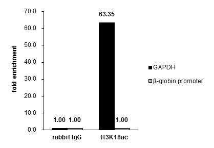
GTX128943 ChIP assay Image
Cross-linked ChIP was performed with HeLa chromatin extract treated with Trichostatin A (0.4 uM for 18 h) and 5 ug of either control rabbit IgG or anti-Histone H3K18ac (acetyl Lys18) antibody. The precipitated DNA was detected by PCR with primer set targeting to GAPDH or ƒÀ-globin promoter.