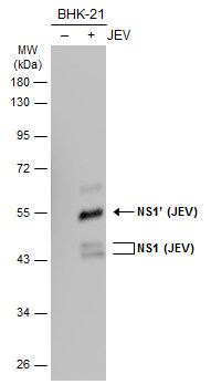
GTX125866 WB Image
Non-infected (?) and infected (+) BHK-21 whole cell extracts (30 ug) were separated by 10% SDS-PAGE, and the membrane was blotted with NS1 (JEV) antibody (GTX125866) diluted at 1:50000. The HRP-conjugated anti-rabbit IgG antibody (GTX213110-01) was used to detect the primary antibody.
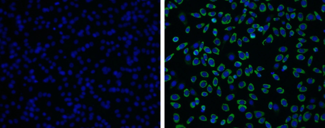
GTX125866 ICC/IF Image
Non-structural protein NS1 (Japanese encephalitis virus) antibody detects non-structural protein NS1 (Japanese encephalitis virus) protein by immunofluorescent analysis.
Samples: BHK-21 cells mock (left) and infected with Japanese encephalitis viruswere fixed in MeOH.
Green: non-structural protein NS1 (Japanese encephalitis virus) protein stained by Non-structural protein NS1 (Japanese encephalitis virus) antibody (GTX125866) diluted at 1:2000.
Blue: Hoechst 33342 staining.
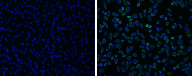
GTX125866 ICC/IF Image
Non-structural protein NS1 (Japanese encephalitis virus) antibody detects non-structural protein NS1 (Japanese encephalitis virus) protein by immunofluorescent analysis.
Samples: BHK-21 cells mock (left) and infected with Japanese encephalitis viruswere fixed in paraformaldehyde.
Green: non-structural protein NS1 (Japanese encephalitis virus) protein stained by Non-structural protein NS1 (Japanese encephalitis virus) antibody (GTX125866) diluted at 1:2000.
Blue: Hoechst 33342 staining.