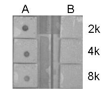
GTX122653 Dot Image
Dotblot analysis of anti-Histone H4K5K8K12K16ac (acetyl Lys5/Lys8/Lys12/Lys16) antibody with peptide samples.
Peptide samples (0.1 ug) were spotted onto positively charged nylon membrane and blotted with Histone H4K5K8K12K16ac (acetyl Lys5/Lys8/Lys12/Lys16) antibody (GTX122653) at different dilution as indicated.
A: Peptide samples of Histone H4K5K8K12K16ac (acetyl Lys5/Lys8/Lys12/Lys16)
B: Peptide samples of Histone H4
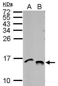
GTX122653 WB Image
Sample (30 ug of whole cell lysate)
A: NIH-3T3
B: BCL-1
15% SDS PAGE
GTX122653 diluted at 1:10000
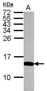
GTX122653 WB Image
Sample (30 ug of whole cell lysate)
A: PC-12
15% SDS PAGE
GTX122653 diluted at 1:10000
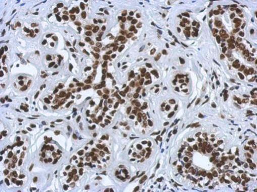
GTX122653 IHC-P Image
Immunohistochemical analysis of paraffin-embedded human breast cancer, using Histone H4K5K8K12K16ac (acetyl Lys5/Lys8/Lys12/Lys16)(GTX122653) antibody at 1:500 dilution.
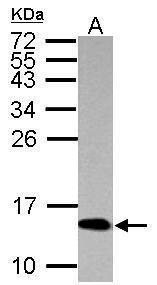
GTX122653 WB Image
Sample (30 ug of whole cell lysate)
A: HeLa
15% SDS PAGE
GTX122653 diluted at 1:10000
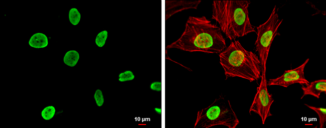
GTX122653 ICC/IF Image
Histone H4K5K8K12K16ac (acetyl Lys5/Lys8/Lys12/Lys16) antibody detects Histone H4K5K8K12K16ac (acetyl Lys5/Lys8/Lys12/Lys16) protein at nucleus by immunofluorescent analysis.
Sample: HeLa cells were fixed in 4% paraformaldehyde at RT for 15 min.
Green: Histone H4K5K8K12K16ac (acetyl Lys5/Lys8/Lys12/Lys16) protein stained by Histone H4K5K8K12K16ac (acetyl Lys5/Lys8/Lys12/Lys16) antibody (GTX122653) diluted at 1:500.
Red: phalloidin, a cytoskeleton marker, diluted at 1:200.
Scale bar = 10 um.