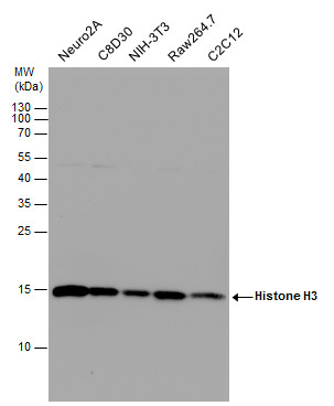
GTX122148 WB Image
Histone H3 antibody detects Histone H3 protein by western blot analysis. Various whole cell extracts (30 ug) were separated by 15% SDS-PAGE, and the membrane was blotted with Histone H3 antibody (GTX122148) diluted at a dilution of 1:10000. The HRP-conjugated anti-rabbit IgG antibody (GTX213110-01) was used to detect the primary antibody.
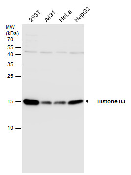
GTX122148 WB Image
Various whole cell extracts (30 ug) were separated by 15% SDS-PAGE, and the membrane was blotted with Histone H3 antibody (GTX122148) diluted at 1:1000. The HRP-conjugated anti-rabbit IgG antibody (GTX213110-01) was used to detect the primary antibody.
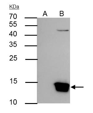
GTX122148 IP Image
Histone H3 antibody immunoprecipitates Histone H3 protein in IP experiments. IP Sample: Raji whole cell lysate/extract A : Control with 3 ug of pre-immune rabbit IgG B : Immunoprecipitation of Histone H3 by 3 ug of Histone H3 antibody (GTX122148) 15% SDS-PAGE The immunoprecipitated Histone H3 protein was detected by Histone H3 antibody (GTX122148) diluted at 1 : 1000. EasyBlot anti-rabbit IgG (HRP) (GTX221666-01) was used as a secondary reagent.
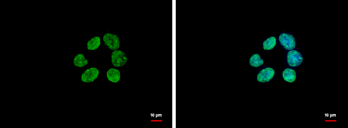
GTX122148 ICC/IF Image
Histone H3 antibody detects Histone H3 protein at nucleus by immunofluorescent analysis.
Sample: A431 cells were fixed in ice-cold MeOH for 5 min.
Green: Histone H3 protein stained by Histone H3 antibody (GTX122148) diluted at 1:500.
Blue: Hoechst 33342 staining.
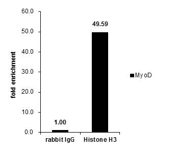
GTX122148 ChIP assay Image
Cross-linked ChIP was performed with HeLa chromatin extract and 5 ug of either control rabbit IgG or anti-Histone H3 antibody. The precipitated DNA was detected by PCR with primer set targeting to MyoD.
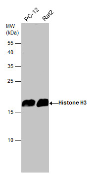
GTX122148 WB Image
Various whole cell extracts (30 ug) were separated by 15% SDS-PAGE, and the membrane was blotted with Histone H3 antibody (GTX122148) diluted at 1:3000. The HRP-conjugated anti-rabbit IgG antibody (GTX213110-01) was used to detect the primary antibody.
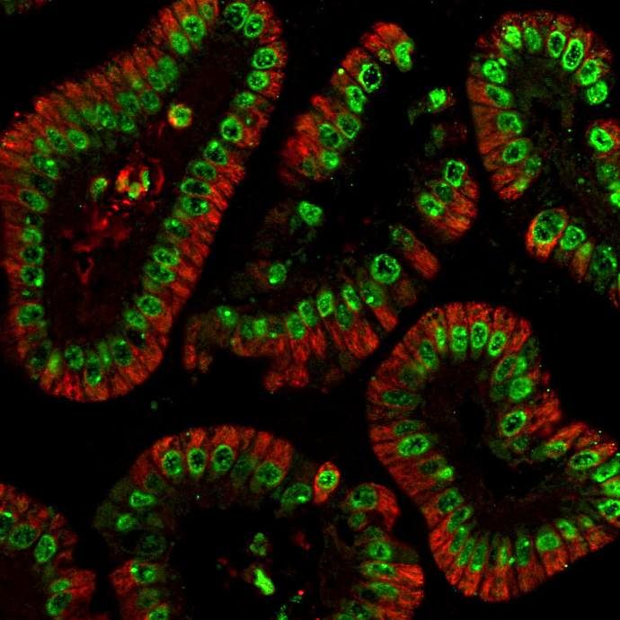
GTX122148 IHC-P Image
Histone H3 antibody detects Histone H3 protein at nucleus in mouse colon by immunohistochemical analysis.
Sample: Paraffin-embedded mouse colon.
Green: Histone H3 antibody (GTX122148) diluted at 1:500.
Red: alpha Tubulin antibody [GT114] (GTX628802) diluted at 1:500.
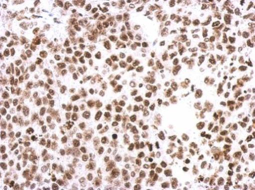
GTX122148 IHC-P Image
Immunohistochemical analysis of paraffin-embedded Hela xenograft, using Histone H3(GTX122148) antibody at 1:500 dilution.
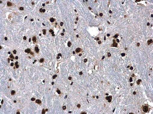
GTX122148 IHC-P Image
Histone H3 antibody detects Histone H3 protein at nucleus in mouse brain by immunohistochemical analysis.
Sample: Paraffin-embedded mouse brain.
Histone H3 antibody (GTX122148) diluted at 1:500.
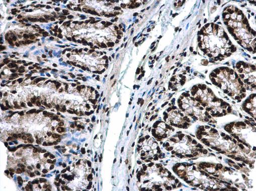
GTX122148 IHC-P Image
Histone H3 antibody detects Histone H3 protein at nucleus in mouse colon by immunohistochemical analysis.
Sample: Paraffin-embedded mouse colon.
Histone H3 antibody (GTX122148) diluted at 1:500.