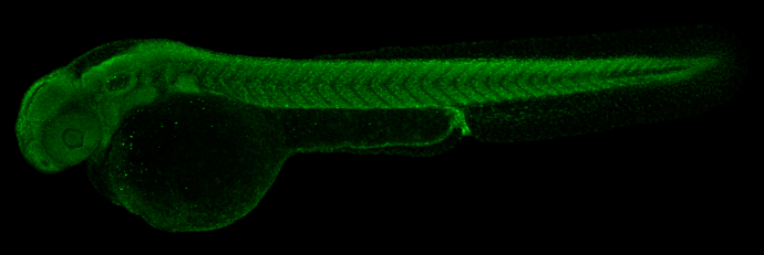
GTX121184 IHC-Wm Image
Histone H3K27me3 (trimethyl Lys27) antibody detects Histone H3K27me3 (trimethyl Lys27) protein on zebrafish by whole mount immunohistochemical analysis. Sample: Paraformaldehyde-fixed 2 day-post-fertilization zebrafish embryo.Histone H3K27me3 (trimethyl Lys27) antibody (GTX121184) dilution: 1:100.
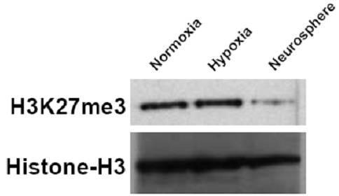
GTX121184 WB Image
U87 cells were grown under normoxic (DMEM 10% FBS, 16% O2), hypoxic (DMEM 10% FBS, 1% O2), and neurosphere conditions (DMEM/F12, B-27 supplement, growth factor (10ng/ml FGF and 20ng/ml EGF)). Cell lysate were Western blotted for H3K27me3 and Histone-H3 (loading control). The HRP-conjugated anti-rabbit IgG antibody (GTX213110-01) was used to detect the primary antibody.
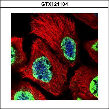
GTX121184 ICC/IF Image
Confocal immunofluorescence analysis (Olympus FV10i) of paraformaldehyde-fixed A431, using Histone H3 (tri-Methyl K27)(GTX121184) antibody (Green) at 1:500 dilution. Alpha-tubulin filaments were labeled with GTX11304 (Red) at 1:2000.
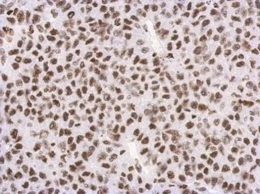
GTX121184 IHC-P Image
Immunohistochemical analysis of paraffin-embedded Hela xenograft, using Histon H3 (tri-Methyl K27)(GTX121184) antibody at 1:500 dilution.
GTX121184 IHC-P Image
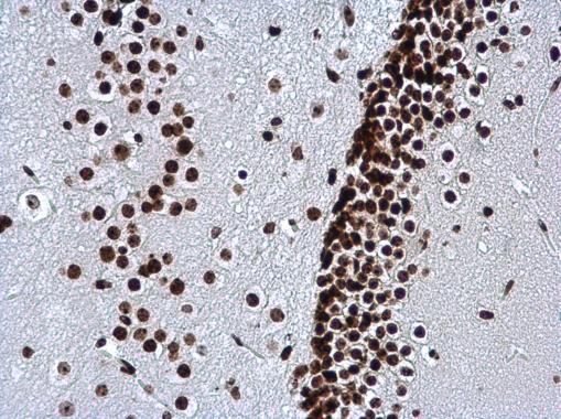
Histone H3K27me3 (trimethyl Lys27) antibody detects Histone H3K27me3 (trimethyl Lys27) protein at nucleus in mouse brain by immunohistochemical analysis.
Sample: Paraffin-embedded mouse brain.
Histone H3K27me3 (trimethyl Lys27) antibody (GTX121184) diluted at 1:500.
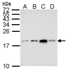
GTX121184 WB Image
Sample (30 ug of whole cell lysate)
A: Jurkat
B: Raji
C: K562
D: THP-1
7.5% SDS PAGE
GTX121184 diluted at 1:500
The HRP-conjugated anti-rabbit IgG antibody (GTX213110-01) was used to detect the primary antibody.
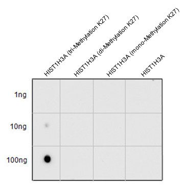
GTX121184 DOT Image
Various peptide samples were spotted onto positively charged nylon membrane, and blotted with Histone H3 (tri-Methyl Lys27) antibody (GTX121184) diluted at 1:2500.
GTX121184 WB Image
Histone H3 (tri-Methyl Lys27) antibody detects Histone H3 (tri-Methyl Lys27) protein by western blot analysis. Various whole cell extracts (30 ug) were separated by 15% SDS-PAGE, and the membrane was blotted with Histone H3 (tri-Methyl Lys27) antibody (GTX121184) diluted at a dilution of 1:1000. The HRP-conjugated anti-rabbit IgG antibody (GTX213110-01) was used to detect the primary antibody.
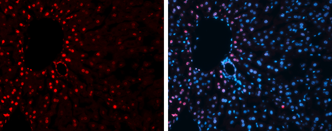
GTX121184 IHC-Fr Image
Immunofluorescence photomicrographs of frozen sections of mouse liver.
Red: Histone H3K27me3 (trimethyl Lys27) antibody (GTX121184) diluted at 1:200. The signal was developed using goat anti-rabbit IgG antibody (Dylight594) (GTX213110-05).
Blue: Nuclear staining with Hoechst 33342.
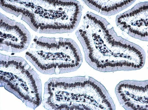
GTX121184 IHC-P Image
Histone H3K27me3 (trimethyl Lys27) antibody detects Histone H3K27me3 (trimethyl Lys27) protein at nucleus on mouse duodenum by immunohistochemical analysis.
Sample: Paraffin-embedded mouse duodenum.
Histone H3K27me3 (trimethyl Lys27) antibody (GTX121184) diluted at 1:500.
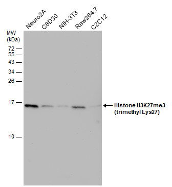
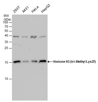
GTX121184 WB Image
Various whole cell extracts (30 ug) were separated by 15% SDS-PAGE, and the membrane was blotted with Histone H3K27me3 (trimethyl Lys27) antibody (GTX121184) diluted at 1:1000.