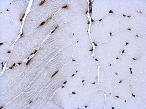
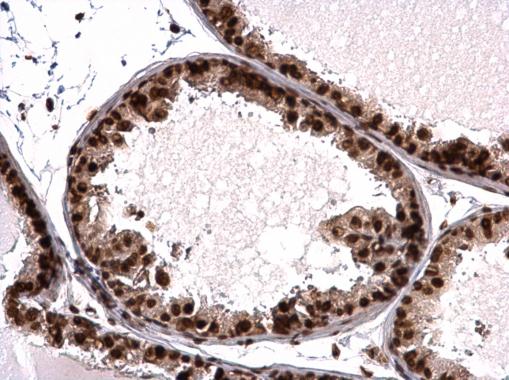
GTX115549 IHC-P Image
Histone H3.3B antibody detects Histone H3.3B protein at nucleus in mouse muscle by immunohistochemical analysis.
Sample: Paraffin-embedded mouse muscle.
Histone H3.3B antibody (GTX115549) diluted at 1:500.
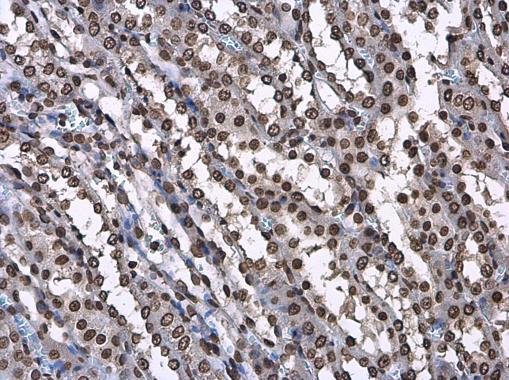
GTX115549 IHC-P Image
Histone H3.3B antibody detects Histone H3.3B protein at nucleus in rat kidney by immunohistochemical analysis.
Sample: Paraffin-embedded rat kidney.
Histone H3.3B antibody (GTX115549) diluted at 1:500.
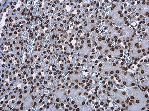
GTX115549 IHC-P Image
Histone H3.3B antibody detects Histone H3.3B protein at nucleus in mouse kidney by immunohistochemical analysis.
Sample: Paraffin-embedded mouse kidney.
Histone H3.3B antibody (GTX115549) diluted at 1:500.
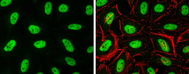
GTX115549 ICC/IF Image
Histone H3.3B antibody detects Histone H3.3B protein at nucleus by immunofluorescent analysis.
Sample: HeLa cells were fixed in 4% paraformaldehyde at RT for 15 min.
Green: Histone H3.3B protein stained by Histone H3.3B antibody (GTX115549) diluted at 1:500.
Red: Phalloidin, a cytoskeleton marker, diluted at 1:100.
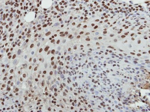
GTX115549 IHC-P Image
Immunohistochemical analysis of paraffin-embedded HSC3 xenograft, using Histone H3.3B(GTX115549) antibody at 1:500 dilution.
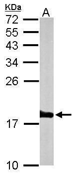
GTX115549 WB Image
Sample (50 ug of whole cell lysate)
A: mouse brain
15% SDS PAGE
GTX115549 diluted at 1:1000
The HRP-conjugated anti-rabbit IgG antibody (GTX213110-01) was used to detect the primary antibody.

GTX115549 WB Image
Histone H3.3B antibody detects H3F3B protein by western blot analysis.
A. 50 ug rat brain lysate/extract
15% SDS-PAGE
Histone H3.3B antibody (GTX115549) dilution: 1:1000
The HRP-conjugated anti-rabbit IgG antibody (GTX213110-01) was used to detect the primary antibody.
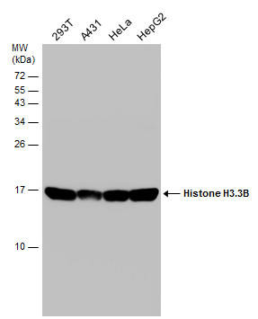
GTX115549 WB Image
Various whole cell extracts (30 ug) were separated by 15% SDS-PAGE, and the membrane was blotted with Histone H3.3B antibody (GTX115549) diluted at 1:10000.
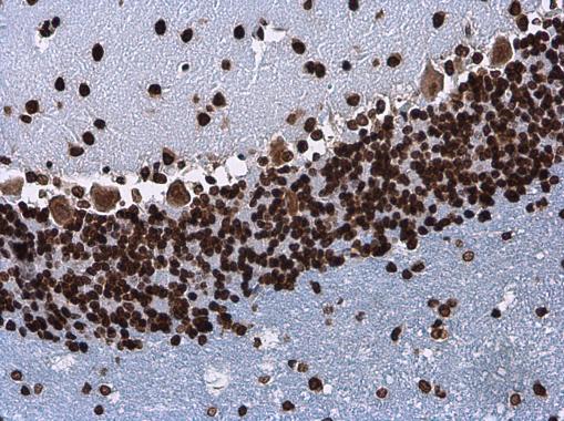
GTX115549 IHC-P Image
Histone H3.3B antibody detects Histone H3.3B protein at nucleus in rat brain by immunohistochemical analysis.
Sample: Paraffin-embedded rat brain.
Histone H3.3B antibody (GTX115549) diluted at 1:500.
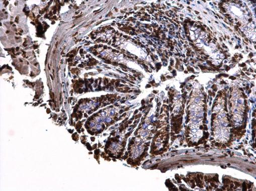
GTX115549 IHC-P Image
Histone H3.3B antibody detects Histone H3.3B protein at nucleus on mouse colon by immunohistochemical analysis.
Sample: Paraffin-embedded mouse colon.
Histone H3.3B antibody (GTX115549) dilution: 1:500.
GTX115549 IHC-P Image
Histone H3.3B antibody detects Histone H3.3B protein at nucleus on mouse prostate by immunohistochemical analysis.
Sample: Paraffin-embedded mouse prostate.
Histone H3.3B antibody (GTX115549) dilution: 1:500.
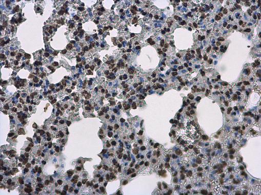
GTX115549 IHC-P Image
Histone H3.3B antibody detects Histone H3.3B protein at nucleus on mouse lung by immunohistochemical analysis.
Sample: Paraffin-embedded mouse lung.
Histone H3.3B antibody (GTX115549) diluted at 1:500.
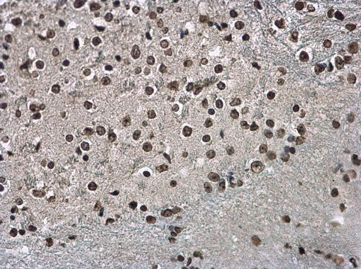
GTX115549 IHC-P Image
Histone H3.3B antibody detects Histone H3.3B protein at nucleus on mouse spinal cord by immunohistochemical analysis.
Sample: Paraffin-embedded mouse spinal cord.
Histone H3.3B antibody (GTX115549) diluted at 1:500.