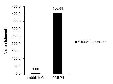
GTX112864 ChIP assay Image
Cross-linked ChIP was performed with Raji chromatin extract and 5 ug of either control rabbit IgG or anti-PARP1 antibody. The precipitated DNA was detected by PCR with primer set targeting to S100A9 promoter.
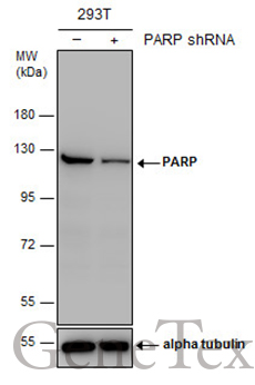
GTX112864 WB Image
Non-transfected (?) and transfected (+) 293T whole cell extracts (30 ug) were separated by 7.5% SDS-PAGE, and the membrane was blotted with PARP antibody [N2C1], Internal (GTX112864) diluted at 1:5000. The HRP-conjugated anti-rabbit IgG antibody (GTX213110-01) was used to detect the primary antibody.
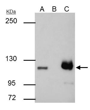
GTX112864 IP Image
PARP1 antibody [N2C1], Internal immunoprecipitates PARP1 protein in IP experiments.
IP samples: HCT-116 whole cell extract
A. 30 ug HCT-116 whole cell extract
B. Control with 4 ug of preimmune Rabbit IgG
C. Immunoprecipitation of PARP1 protein by 4 ug PARP1 antibody [N2C1], Internal (GTX112864)
5 % SDS-PAGE
The immunoprecipitated PARP1 protein was detected by PARP1 antibody [N2C1], Internal (GTX112864) diluted at 1:500.
[EasyBlot anti-rabbit IgG (GTX221666-01) was used as a secondary reagent]
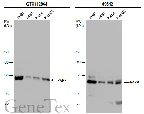
GTX112864 WB Image
Various whole cell extracts (30 ug) were separated by 5% SDS-PAGE, and the membranes were blotted with PARP antibody [N2C1], Internal (GTX112864) diluted at 1:10000 and competitor's antibody (#9542) diluted at 1:500. The HRP-conjugated anti-rabbit IgG antibody (GTX213110-01) was used to detect the primary antibody.
*The competitor is not affiliated with GeneTex and does not endorse this product.
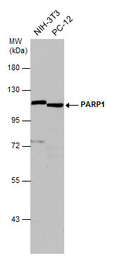
GTX112864 WB Image
Various whole cell extracts (30 ug) were separated by 7.5% SDS-PAGE, and the membrane was blotted with PARP1 antibody [N2C1], Internal (GTX112864) diluted at 1:500. The HRP-conjugated anti-rabbit IgG antibody (GTX213110-01) was used to detect the primary antibody.
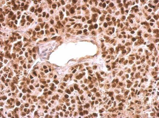
GTX112864 IHC-P Image
PARP1 antibody [N2C1], Internal detects PARP1 protein at nucleus on HeLa xenograft by immunohistochemical analysis.
Sample: Paraffin-embedded HeLa xenograft.
PARP1 antibody [N2C1], Internal (GTX112864) dilution: 1:500.
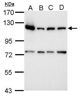
GTX112864 WB Image
Sample (30 ug of whole cell lysate)
A: 293T
B: A431
C: HeLa
D: HepG2
7.5% SDS PAGE
GTX112864 diluted at 1:10000
The HRP-conjugated anti-rabbit IgG antibody (GTX213110-01) was used to detect the primary antibody.
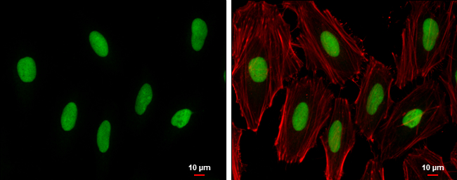
GTX112864 ICC/IF Image
PARP antibody [N2C1], Internal detects PARP protein at nucleus by immunofluorescent analysis.
Sample: HeLa cells were fixed in 4% paraformaldehyde at RT for 15 min.
Green: PARP protein stained by PARP antibody [N2C1], Internal (GTX112864) diluted at 1:500.
Red: phalloidin, a cytoskeleton marker, diluted at 1:200.
Blue: Hoechst 33342 staining.
Scale bar = 10 um.
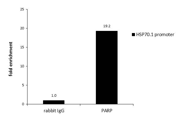
GTX112864 ChIP assay Image
ChIP was performed with HeLa chromatin extract and 5 ug of either normal rabbit IgG or anti-PARP antibody. The precipitated DNA was detected by PCR with primer set targeting to HSP70.1 promoter.
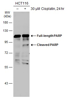
GTX112864 WB Image
Untreated (?) and treated (+) HCT-116 whole cell extract (30 ug) were separated by 7.5% SDS-PAGE, and the membrane was blotted with PARP antibody (GTX112864) diluted at 1:1000. The HRP-conjugated anti-rabbit IgG antibody (GTX213110-01) was used to detect the primary antibody.