
GTX111506 IHC-P Image
HKDC1 antibody detects HKDC1 protein at cytoplasm in mouse duodenum by immunohistochemical analysis.
Sample: Paraffin-embedded mouse duodenum.
HKDC1 antibody (GTX111506) diluted at 1:500.

GTX111506 IHC-P Image
HKDC1 antibody detects HKDC1 protein at cytoplasm in rat brain by immunohistochemical analysis.
Sample: Paraffin-embedded rat brain.
HKDC1 antibody (GTX111506) diluted at 1:500.
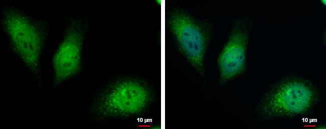
GTX111506 ICC/IF Image
HKDC1 antibody detects HKDC1 protein at cytoplasm and nucleus by immunofluorescent analysis.
Sample: HeLa cells were fixed in 4% paraformaldehyde at RT for 15 min.
Green: HKDC1 protein stained by HKDC1 antibody (GTX111506) diluted at 1:500.
Blue: Hoechst 33342 staining.

GTX111506 WB Image
Whole cell extract (30 ug) was separated by 7.5% SDS-PAGE, and the membrane was blotted with HKDC1 antibody (GTX111506) diluted at 1:2000.