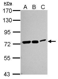
GTX111069 WB Image
Sample (30 ug of whole cell lysate)
A: Neuro2A
B: GL261
C: C8D30
7.5% SDS PAGE
GTX111069 diluted at 1:1000
The HRP-conjugated anti-rabbit IgG antibody (GTX213110-01) was used to detect the primary antibody.
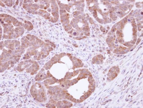
GTX111069 IHC-P Image
Immunohistochemical analysis of paraffin-embedded NCIN87 xenograft, using HSC70(GTX111069) antibody at 1:100 dilution.
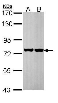
GTX111069 WB Image
Sample (30 ug of whole cell lysate)
A: A431 (GTX27909)
B: HeLa
7.5% SDS PAGE
GTX111069 diluted at 1:1000
The HRP-conjugated anti-rabbit IgG antibody (GTX213110-01) was used to detect the primary antibody.
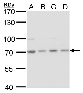
GTX111069 WB Image
HSC70 antibody detects HSC70 protein by western blot analysis.
A. 30 ug 293T whole cell lysate/extract
B. 30 ug A431whole cell lysate/extract
C. 30 ug HeLa whole cell lysate/extract
D. 30 ug HepG2 whole cell lysate/extract
7.5% SDS-PAGE
HSC70 antibody (GTX111069) dilution: 1:1000
The HRP-conjugated anti-rabbit IgG antibody (GTX213110-01) was used to detect the primary antibody.
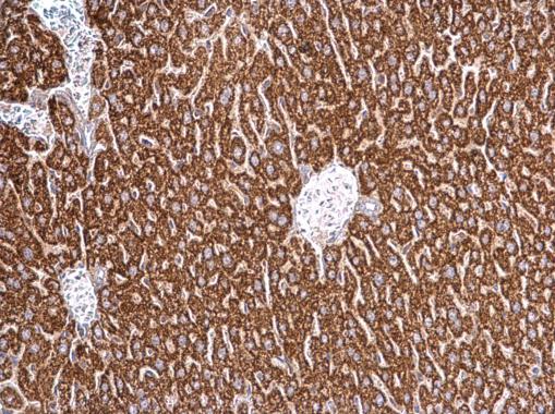
GTX111069 IHC-P Image
HSC70 antibody detects HSC70 protein at cytosol on mouse liver by immunohistochemical analysis.
Sample: Paraffin-embedded mouse liver.
HSC70 antibody (GTX111069) dilution: 1:500.
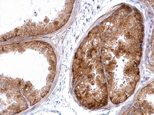
GTX111069 IHC-P Image
HSC70 antibody detects HSC70 protein at cytosol on mouse prostate by immunohistochemical analysis.
Sample: Paraffin-embedded mouse prostate.
HSC70 antibody (GTX111069) dilution: 1:500.
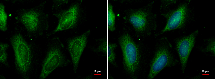
GTX111069 ICC/IF Image
HSC70 antibody detects HSC70 protein at cytoplasm by immunofluorescent analysis.
Sample: HeLa cells were fixed in ice-cold MeOH for 5 min.
Green: HSC70 protein stained by HSC70 antibody (GTX111069) diluted at 1:500.
Blue: Hoechst 33342 staining.