GTX110564 WB Image
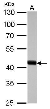
beta Actin antibody detects ACTB protein by western blot analysis.
A. 30 ug drosophila lysate/extract
10% SDS-PAGE
beta Actin antibody (GTX110564) dilution: 1:10000
The HRP-conjugated anti-rabbit IgG antibody (GTX213110-01) was used to detect the primary antibody.
GTX110564 IHC-P Image
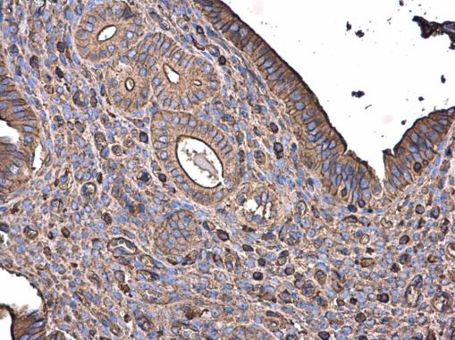
beta Actin antibody detects beta Actin protein at cytoplasm in mouse cervix by immunohistochemical analysis.
Sample: Paraffin-embedded mouse cervix.
beta Actin antibody (GTX110564) diluted at 1:500.
GTX110564 WB Image
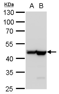
beta Actin antibody detects beta Actin protein by western blot analysis.
A. 30 ug PC-12 whole cell lysate/extract
B. 30 ug Rat2 whole cell lysate/extract
10% SDS-PAGE
beta Actin antibody (GTX110564) dilution: 1:20000
The HRP-conjugated anti-rabbit IgG antibody (GTX213110-01) was used to detect the primary antibody.
GTX110564 IP Image
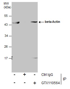
Immunoprecipitation of beta Actin protein from 293T whole cell extracts using 5 ug of beta Actin antibody (GTX110564).
Western blot analysis was performed using beta Actin antibody (GTX110564).
EasyBlot anti-Rabbit IgG (GTX221666-01) was used as a secondary reagent.
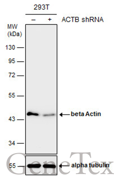
GTX110564 WB Image
Non-transfected (?) and transfected (+) 293T whole cell extracts (10 ug) were separated by 10% SDS-PAGE, and the membrane was blotted with beta Actin antibody (GTX110564) diluted at 1:15000. The HRP-conjugated anti-rabbit IgG antibody (GTX213110-01) was used to detect the primary antibody.
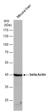
GTX110564 WB Image
beta Actin antibody detects beta Actin protein by western blot analysis. Mouse tissue extracts (50 ug) was separated by 10% SDS-PAGE, and the membrane was blotted with beta Actin antibody (GTX110564) diluted by 1:20000. The HRP-conjugated anti-rabbit IgG antibody (GTX213110-01) was used to detect the primary antibody.
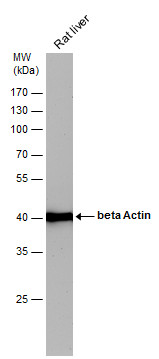
GTX110564 WB Image
beta Actin antibody detects beta Actin protein by western blot analysis. Rat tissue extracts (50 ug) was separated by 10% SDS-PAGE, and the membrane was blotted with beta Actin antibody (GTX110564) diluted by 1:20000. The HRP-conjugated anti-rabbit IgG antibody (GTX213110-01) was used to detect the primary antibody.
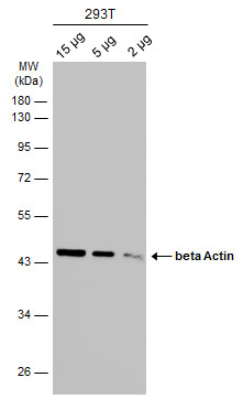
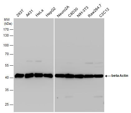
GTX110564 WB Image
Various whole cell extracts (15
2 ug) were separated by 10% SDS-PAGE, and the membrane was blotted with beta Actin antibody (GTX110564) diluted at 1:10000. The HRP-conjugated anti-rabbit IgG antibody (GTX213110-01) was used to detect the primary antibody.
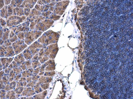
GTX110564 IHC-P Image
beta Actin antibody detects beta Actin protein at cell membrane and cytoplasm in mouse pancreas by immunohistochemical analysis.
Sample: Paraffin-embedded mouse pancreas.
beta Actin antibody (GTX110564) diluted at 1:500.
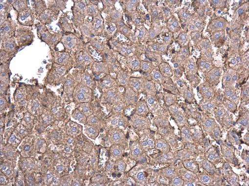
GTX110564 IHC-P Image
beta Actin antibody detects beta Actin protein at cell membrane and cytoplasm in rat liver by immunohistochemical analysis.
Sample: Paraffin-embedded rat liver.
beta Actin antibody (GTX110564) diluted at 1:500.
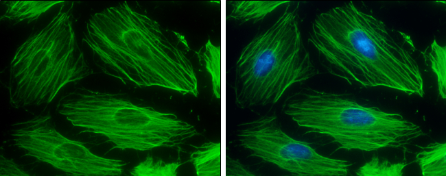
GTX110564 ICC/IF Image
beta Actin antibody detects beta Actin protein at cytoskeleton by immunofluorescent analysis.
Sample: HeLa cells were fixed in 0.5% Triton X-100 for 1 min, then ice-cold methanol for 5 min.
Green: beta Actin protein stained by beta Actin antibody (GTX110564) diluted at 1:500.
Blue: Hoechst 33342 staining.
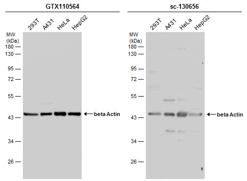
GTX110564 WB Image
Various whole cell extracts (30 ug) were separated by 10% SDS-PAGE, and the membranes were blotted with beta Actin antibody (GTX110564) diluted at 1:10000 and competitor's antibody (sc-130656) diluted at 1:100. The HRP-conjugated anti-rabbit IgG antibody (GTX213110-01) was used to detect the primary antibody.
GTX110564 WB Image
beta Actin antibody detects beta Actin protein by western blot analysis. Various whole cell extracts (30 ug) were separated by 10% SDS-PAGE, and the membrane was blotted with beta Actin antibody (GTX110564) diluted at a dilution of 1:20000. The HRP-conjugated anti-rabbit IgG antibody (GTX213110-01) was used to detect the primary antibody.
