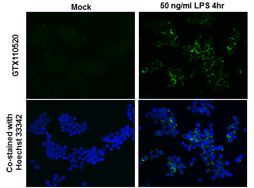
GTX110520 ICC/IF Image
TNF alpha antibody detects TNF alpha protein at cytoplasm by immunofluorescent analysis.
Sample: Raw264.7 cells were fixed in 4% paraformaldehyde at RT for 15 min.
Green: TNF alpha protein stained by TNF alpha antibody (GTX110520) diluted at 1:500.
Blue: Hoechst 33342 staining.
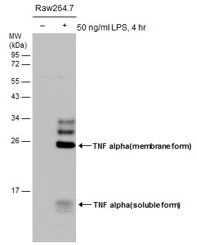
GTX110520 WB Image
Untreated (?) and treated (+) Raw264.7 whole cell extracts (30 ug) were separated by 12% SDS-PAGE, and the membrane was blotted with TNF alpha antibody (GTX110520) diluted at 1:1000.
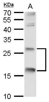
GTX110520 WB Image
TNF alpha antibody detects TNF alpha protein by western blot analysis.
A. 30 ug NCI-H929 whole cell lysate/extract
12% SDS-PAGE
TNF alpha antibody (GTX110520) dilution: 1:500
The HRP-conjugated anti-rabbit IgG antibody (GTX213110-01) was used to detect the primary antibody.
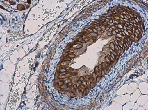
GTX110520 IHC-P Image
TNF alpha antibody detects TNF alpha protein at cell membrane in mouse prostate by immunohistochemical analysis.
Sample: Paraffin-embedded mouse prostate.
TNF alpha antibody (GTX110520) diluted at 1:400.
GTX110520 IHC-P Image
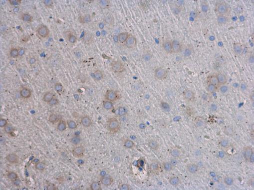
TNF alpha antibody detects TNF alpha protein at cell membrane in rat brain by immunohistochemical analysis.
Sample: Paraffin-embedded rat brain.
TNF alpha antibody (GTX110520) diluted at 1:400.
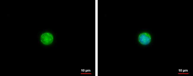
GTX110520 ICC/IF Image
TNF alpha antibody detects TNF alpha protein at membrane by immunofluorescent analysis.
Sample: jurkat cells were fixed in 4% paraformaldehyde at RT for 15 min.
Green: TNF alpha protein stained by TNF alpha antibody (GTX110520) diluted at 1:500.
Blue: Hoechst 33342 staining.
GTX110520 WB Image
Whole cell extract (30 ug) was separated by 12% SDS-PAGE, and the membrane was blotted with TNF alpha antibody (GTX110520) diluted at 1:1000. The HRP-conjugated anti-rabbit IgG antibody (GTX213110-01) was used to detect the primary antibody.
GTX110520 IHC-P Image
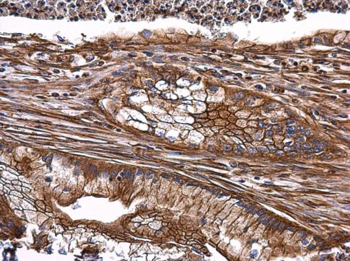
TNF alpha antibody detects TNF alpha protein at cytoplasm on human esophagus carcinoma by immunohistochemical analysis.
Sample: Paraffin-embedded human esophagus carcinoma .
TNF alpha antibody (GTX110520) diluted at 1:500.
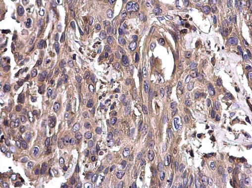
GTX110520 IHC-P Image
TNF alpha antibody detects TNF alpha protein at membrane on human gastric carcinoma by immunohistochemical analysis.
Sample: Paraffin-embedded human gastric carcinoma.
TNF alpha antibody (GTX110520) dilution: 1:500.
GTX110520 WB Image
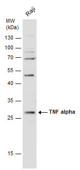
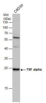
TNF alpha antibody detects TNF alpha protein by western blot analysis. Whole cell extracts (30 ug) was separated by 12% SDS-PAGE, and the membrane was blotted with TNF alpha antibody (GTX110520) diluted at 1:500. The HRP-conjugated anti-rabbit IgG antibody (GTX213110-01) was used to detect the primary antibody.
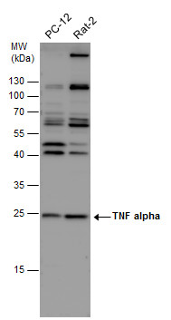
GTX110520 WB Image
TNF alpha antibody detects TNF alpha protein by western blot analysis. Various whole cell extracts (30 ug) were separated by 12% SDS-PAGE, and the membrane was blotted with TNF alpha antibody (GTX110520) diluted at 1:500. The HRP-conjugated anti-rabbit IgG antibody (GTX213110-01) was used to detect the primary antibody.