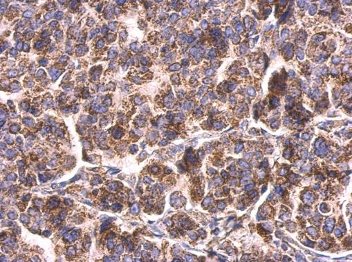
GTX110089 IHC-P Image
HSP60 antibody detects HSP60 protein at mitochondria on human breast carcinoma by immunohistochemical analysis.
Sample: Paraffin-embedded human breast carcinoma.
HSP60 antibody (GTX110089) dilution: 1:500.
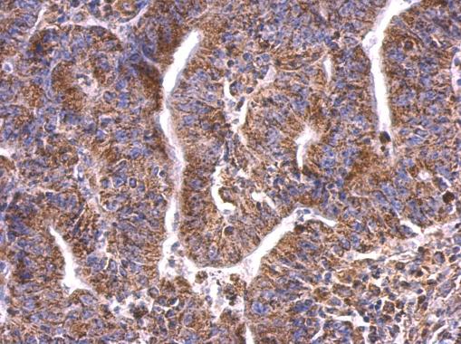
GTX110089 IHC-P Image
HSP60 antibody detects HSP60 protein at mitochondria on human colon carcinoma by immunohistochemical analysis.
Sample: Paraffin-embedded human colon carcinoma.
HSP60 antibody (GTX110089) dilution: 1:500.
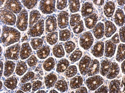
GTX110089 IHC-P Image
HSP60 antibody detects HSP60 protein at mitochondrion on mouse intestine by immunohistochemical analysis.
Sample: Paraffin-embedded mouse intestine.
HSP60 antibody (GTX110089) dilution: 1:500.
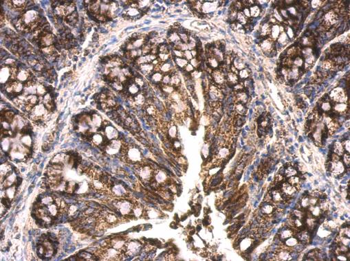
GTX110089 IHC-P Image
HSP60 antibody detects HSP60 protein at mitochondrion on mouse colon by immunohistochemical analysis.
Sample: Paraffin-embedded mouse colon.
HSP60 antibody (GTX110089) dilution: 1:500.
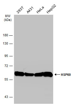
GTX110089 WB Image
Various whole cell extracts (30 ug) were separated by 7.5% SDS-PAGE, and the membrane was blotted with HSP60 antibody (GTX110089) diluted at 1:10000. The HRP-conjugated anti-rabbit IgG antibody (GTX213110-01) was used to detect the primary antibody.
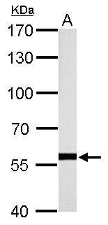
GTX110089 WB Image
HSP60 antibody detects HSPD1 protein by western blot analysis.
A. 50 ug rat brain lysate/extract
7.5% SDS-PAGE
HSP60 antibody (GTX110089) dilution: 1:10000
The HRP-conjugated anti-rabbit IgG antibody (GTX213110-01) was used to detect the primary antibody.
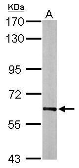
GTX110089 WB Image
Sample (20 ug of whole cell lysate)
A: mouse brain
7.5% SDS PAGE
GTX110089 diluted at 1:20000
The HRP-conjugated anti-rabbit IgG antibody (GTX213110-01) was used to detect the primary antibody.
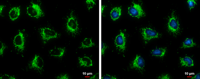
GTX110089 ICC/IF Image
HSP60 antibody detects HSP60 protein at mitochondria by immunofluorescent analysis.
Sample: HeLa cells were fixed in ice-cold MeOH for 5 min.
Green: HSP60 protein stained by HSP60 antibody (GTX110089) diluted at 1:500.
Blue: Hoechst 33342 staining.
Scale bar = 10 um.