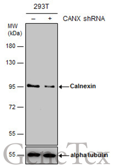
GTX109669 WB Image
Non-transfected (?) and transfected (+) 293T whole cell extracts (15 ug) were separated by 7.5% SDS-PAGE, and the membrane was blotted with Calnexin antibody [C3], C-term (GTX109669) diluted at 1:20000. The HRP-conjugated anti-rabbit IgG antibody (GTX213110-01) was used to detect the primary antibody.
GTX109669 WB Image
Calnexin antibody [C3], C-term detects Calnexin protein by Western blot analysis.
A. 30 ug Neuro2A whole cell lysate/extract
B. 30 ug GL261 whole cell lysate/extract
C. 30 ug C8D30 whole cell lysate/extract
D. 30 ug NIH-3T3 whole cell lysate/extract
E. 30 ug BCL-1 whole cell lysate/extract
F. 30 ug Raw264.7 whole cell lysate/extract
G. 30 ug C2C12 whole cell lysate/extract
7.5 % SDS-PAGE
Calnexin antibody [C3], C-term (GTX109669) dilution: 1:10000

GTX109669 IHC-P Image
Calnexin antibody [C3], C-term detects Calnexin protein at cytosol on mouse middle brain by immunohistochemical analysis.
Sample: Paraffin-embedded mouse middle brain.
Calnexin antibody [C3], C-term (GTX109669) dilution: 1:500.

GTX109669 IHC-P Image
Calnexin antibody [C3], C-term detects Calnexin protein at cytosol on mouse testis by immunohistochemical analysis.
Sample: Paraffin-embedded mouse testis.
Calnexin antibody [C3], C-term (GTX109669) dilution: 1:500.
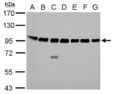
GTX109669 WB Image
Various whole cell extracts (30 ug) were separated by 7.5% SDS-PAGE, and the membrane was blotted with Calnexin antibody [C3], C-term (GTX109669) diluted at 1:10000. The HRP-conjugated anti-rabbit IgG antibody (GTX213110-01) was used to detect the primary antibody.

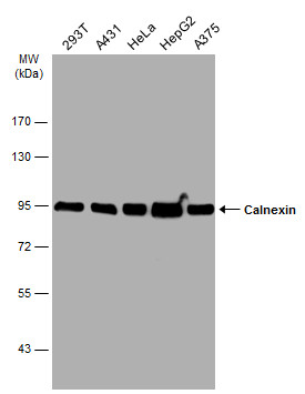
GTX109669 WB Image
Various whole cell extracts (30 ug) were separated by 7.5% SDS-PAGE, and the membrane was blotted with Calnexin antibody [C3], C-term (GTX109669) diluted at 1:10000. The HRP-conjugated anti-rabbit IgG antibody (GTX213110-01) was used to detect the primary antibody.

GTX109669 IP Image
Calnexin antibody immunoprecipitates Calnexin protein in IP experiments. IP Sample: 1000 ug HeLa whole cell lysate/extract A. 30 ug HeLa whole cell lysate/extract B. Control with 2 ug of preimmune rabbit IgG C. Immunoprecipitation of Calnexin protein by 2 ug of Calnexin antibody (GTX109669) 7.5% SDS-PAGE The immunoprecipitated Calnexin protein was detected by Calnexin antibody (GTX109669) diluted at 1:1000. EasyBlot anti-rabbit IgG (GTX221666-01) was used as a secondary reagent.
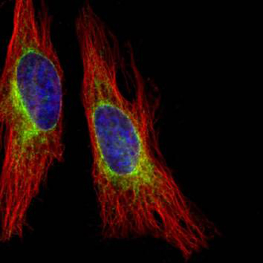
GTX109669 ICC/IF Image
Confocal immunofluorescence analysis (Olympus FV10i) of methanol-fixed HeLa, using Calnexin(GTX109669) antibody (Green) at 1:500 dilution. Alpha-tubulin filaments were labeled with GTX11304 (Red) at 1:2500.
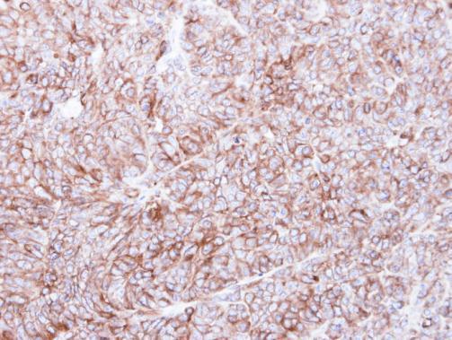
GTX109669 IHC-P Image
Immunohistochemical analysis of paraffin-embedded DLD-1 xenograft, using Calnexin(GTX109669) antibody at 1:500 dilution.
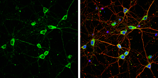
GTX109669 ICC/IF Image
Calnexin antibody [C3], C-term detects Calnexin protein by immunofluorescent analysis.Sample: DIV9 rat E18 primary cortical neuron cells were fixed in 4% paraformaldehyde at RT for 15 min.Green: Calnexin stained by Calnexin antibody [C3], C-term (GTX109669) diluted at 1:500.Red: beta Tubulin 3/ Tuj1, stained by beta Tubulin 3/ Tuj1 antibody [GT1338] (GTX631831) diluted at 1:500.Blue: Fluoroshield with DAPI (GTX30920).