
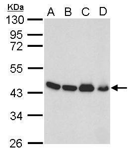
GTX109639 WB Image
Sample (30 ug of whole cell lysate)
A: 293T
B: NTH-3T3
C: mouse brain
D: PC-12
E: Rat brain
F: drosophila
10% SDS PAGE
GTX109639 diluted at 1:10000
The HRP-conjugated anti-rabbit IgG antibody (GTX213110-01) was used to detect the primary antibody.
GTX109639 WB Image
Sample (whole cell lysate)
A: 293T 20ug
B: 293T 10ug
C: 293T 5ug
D: 293T 1ug
10% SDS PAGE
GTX109639 diluted at 1:5000
The HRP-conjugated anti-rabbit IgG antibody (GTX213110-01) was used to detect the primary antibody.
GTX109639 WB Image
Sample (10 ug of whole cell lysate)
A: Yeast lysate
7.5% SDS PAGE
GTX109639 diluted at 1:1000
The HRP-conjugated anti-rabbit IgG antibody (GTX213110-01) was used to detect the primary antibody.
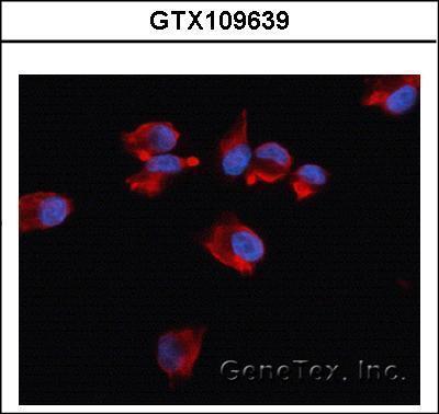
GTX109639 ICC/IF Image
Immunofluorescence analysis of methanol-fixed HeLa, using Beta-actin (GTX109639) antibody at 1:500 dilution.
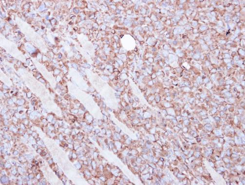
GTX109639 IHC-P Image
Immunohistochemical analysis of paraffin-embedded H1299 xenograft, using Beta actin(GTX109639) antibody at 1:100 dilution.
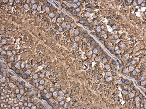
GTX109639 IHC-P Image
beta Actin antibody detects beta Actin protein at cytoplasm in rat testis by immunohistochemical analysis.
Sample: Paraffin-embedded rat testis.
beta Actin antibody (GTX109639) diluted at 1:500.
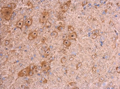
GTX109639 IHC-P Image
beta Actin antibody detects beta Actin protein at cytoplasm in mouse brain by immunohistochemical analysis.
Sample: Paraffin-embedded mouse brain.
beta Actin antibody (GTX109639) diluted at 1:500.
GTX109639 WB Image
Sample (30 ug of whole cell lysate)
A: 293T
B: A431
C: Jurkat
D: Raji
10% SDS PAGE
GTX109639 diluted at 1:1000
The HRP-conjugated anti-rabbit IgG antibody (GTX213110-01) was used to detect the primary antibody.
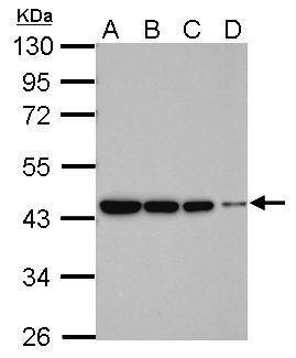
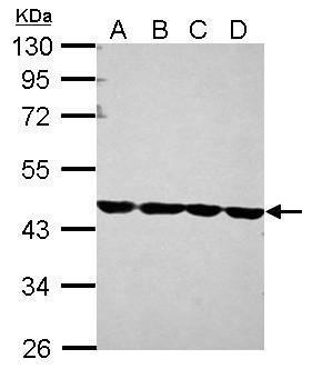
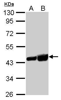
GTX109639 WB Image
beta Actin antibody detects ACTB protein by western blot analysis.
A. 50 ug mouse brain lysate/extract
B. 50 ug mouse kidney lysate/extract
C. 50 ug mouse lung lysate/extract
D. 50 ug mouse testis lysate/extract
10% SDS-PAGE
beta Actin antibody (GTX109639) dilution: 1:10000
The HRP-conjugated anti-rabbit IgG antibody (GTX213110-01) was used to detect the primary antibody.
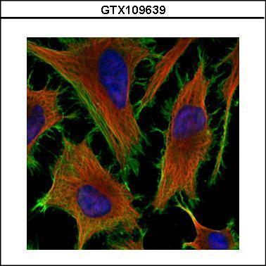
GTX109639 ICC/IF Image
Confocal immunofluorescence analysis (Olympus FV10i) of methanol-fixed HeLa, using beta Actin(GTX109639) antibody (Green) at 1:200 dilution. Alpha-tubulin filaments were labeled with GTX11304 (Red) at 1:2000.
GTX109639 WB Image
Sample (whole cell lysate)
A: Candida albicans (sc5314) 20ug
B: Candida albicans (sc5314) 40ug
10% SDS PAGE
GTX109639 diluted at 1:4000
The HRP-conjugated anti-rabbit IgG antibody (GTX213110-01) was used to detect the primary antibody.

GTX109639 WB Image
Sample (30 ug of whole cell lysate)
A: Jurkat
B: Raji
C: 293T
D: A431
E: HeLa
F: HepG2
G: H1299
H: HCT116
I: MCF-7
J: NT2D1
K: PC-3
L: U87-MG
10% SDS PAGE
GTX109639 diluted at 1:10000
The HRP-conjugated anti-rabbit IgG antibody (GTX213110-01) was used to detect the primary antibody.
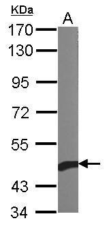
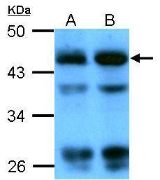
GTX109639 WB Image
beta Actin antibody detects ACTB protein by western blot analysis.
A. 30 ug PC-12 whole cell lysate/extract
B. 30 ug Rat-2 whole cell lysate/extract
10% SDS-PAGE
beta Actin antibody (GTX109639) dilution: 1:5000
The HRP-conjugated anti-rabbit IgG antibody (GTX213110-01) was used to detect the primary antibody.
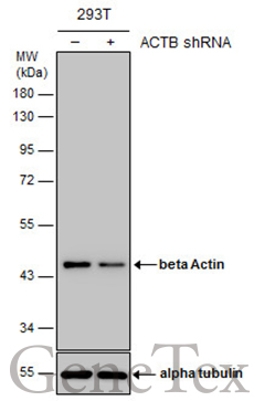
GTX109639 WB Image
Non-transfected (?) and transfected (+) 293T whole cell extracts (10 ug) were separated by 10% SDS-PAGE, and the membrane was blotted with beta Actin antibody (GTX109639) diluted at 1:15000. The HRP-conjugated anti-rabbit IgG antibody (GTX213110-01) was used to detect the primary antibody.