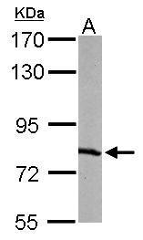
GTX108749 WB Image
Sample (50 ug of whole cell lysate)
A: mouse liver
7.5% SDS PAGE
GTX108749 diluted at 1:1000
The HRP-conjugated anti-rabbit IgG antibody (GTX213110-01) was used to detect the primary antibody.
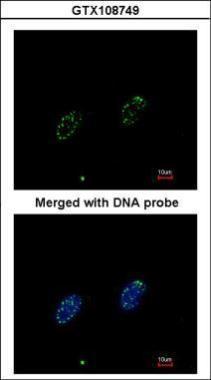
GTX108749 ICC/IF Image
Immunofluorescence analysis of paraformaldehyde-fixed HeLa, using RPA 70 kDa subunit (GTX108749) antibody at 1:200 dilution.
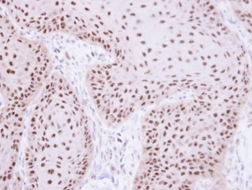
GTX108749 IHC-P Image
Immunohistochemical analysis of paraffin-embedded Cal27 xenograft, using RPA 70 kDa subunit (GTX108749) antibody at 1:100 dilution.
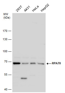
GTX108749 WB Image
RPA70 antibody detects RPA70 protein by western blot analysis. Various whole cell extracts (30 ug) were separated by 7.5% SDS-PAGE, and the membrane was blotted with RPA70 antibody (GTX108749) diluted by 1:1000. The HRP-conjugated anti-rabbit IgG antibody (GTX213110-01) was used to detect the primary antibody.
GTX108749 IHC-P Image
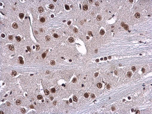
RPA70 antibody [C1C3] detects RPA1 protein at nucleus in mouse brain by immunohistochemical analysis.
Sample: Paraffin-embedded mouse brain.
RPA70 antibody [C1C3] (GTX108749) diluted at 1:500.
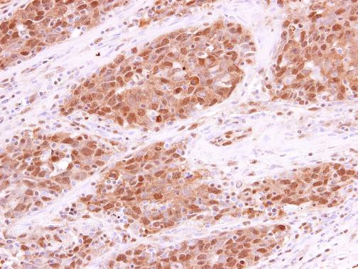
GTX108749 IHC-P Image
RPA70 antibody [C1C3] detects RPA70 protein at cytoplasm and nucleus in human lung papillary adenocarcinoma by immunohistochemical analysis.
Sample: Paraffin-embedded human lung papillary adenocarcinoma.
RPA70 antibody [C1C3] (GTX108749) diluted at 1:250.
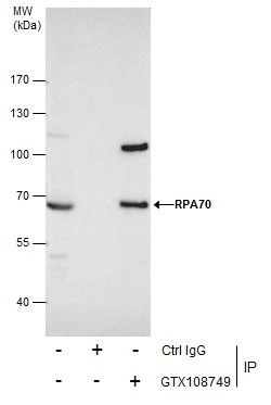
GTX108749 IP Image
Immunoprecipitation of RPA70 protein from 293T whole cell extracts using 5 ug of RPA70 antibody [C1C3] (GTX108749).
Western blot analysis was performed using RPA70 antibody [C1C3] (GTX108749).
EasyBlot anti-Rabbit IgG (GTX221666-01) was used as a secondary reagent.