
GTX102175 IHC-P Image
PAF49 antibody detects PAF49 protein at cytoplasm and nucleus in human ovarian cancer by immunohistochemical analysis.
Sample: Paraffin-embedded human ovarian cancer.
PAF49 antibody (GTX102175) diluted at 1:500.

GTX102175 ICC/IF Image
PAF49 antibody detects PAF49 protein at nucleolus by immunofluorescent analysis.
Sample: HeLa cells were fixed in ice-cold MeOH for 5 min.
Green: PAF49 protein stained by PAF49 antibody (GTX102175) diluted at 1:1000.
Red: alpha Tubulin, a cytoskeleton marker, stained by alpha Tubulin antibody [GT114] (GTX628802) diluted at 1:500.
Blue: Hoechst 33342 staining.
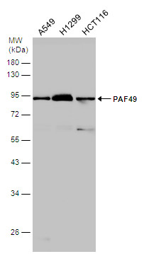
GTX102175 WB Image
Various whole cell extracts (30 ug) were separated by 10% SDS-PAGE, and the membrane was blotted with PAF49 antibody (GTX102175) diluted at 1:1000.
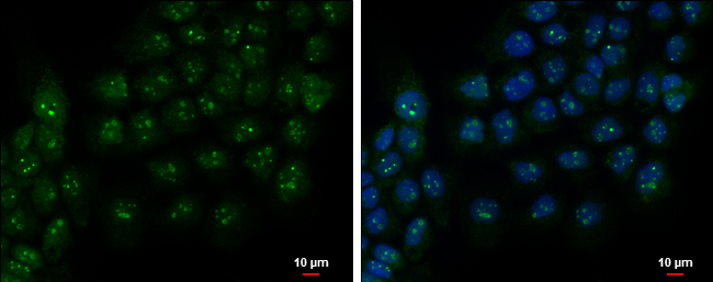
GTX102175 ICC/IF Image
PAF49 antibody detects PAF49 protein at nucleolus by immunofluorescent analysis.
Sample: A431 cells were fixed in 4% paraformaldehyde at RT for 15 min.
Green: PAF49 protein stained by PAF49 antibody (GTX102175) diluted at 1:500.
Blue: Hoechst 33342 staining.
Scale bar = 10 um.