
GTX101766 IHC-P Image
Immunohistochemical analysis of paraffin-embedded human lung Papillary adenocarcinoma, using CD81(GTX101766) antibody at 1:250 dilution.

GTX101766 WB Image
Non-transfected (?) and transfected (+) 293T whole cell extracts (30 ug) were separated by 12% SDS-PAGE, and the membrane was blotted with CD81 antibody (GTX101766) diluted at 1:1000.
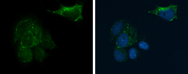
GTX101766 ICC/IF Image
CD81 antibody detects CD81 protein at cell membrane and cytoplasm by immunofluorescent analysis.
Sample: MCF7 cells were fixed in 4% paraformaldehyde at RT for 15 min.
Green: CD81 protein stained by CD81 antibody (GTX101766) diluted at 1:500.
Blue: Hoechst 33342 staining.

GTX101766 FACS Image
CD81 antibody (GTX101766) detects CD81 protein by flow cytometry analysis.
Sample: THP-1 cell.
Black: Unlabelled sample was used as a control.
Red: CD81 antibody (GTX101766) dilution: 1:50.
The Rabbit IgG antibody (DyLight488) (GTX213110-04) was used to detect the primary antibody.