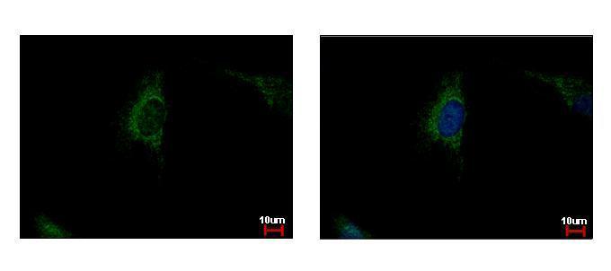
GTX101689 ICC/IF Image
SDHA antibody detects SDHA protein at mitochondria by immunofluorescent analysis.
Sample: HeLa cells were fixed in 4% paraformaldehyde at RT for 15 min.
Green: SDHA protein stained by SDHA antibody (GTX101689) diluted at 1:500.
Blue: Hoechst 33343 staining.
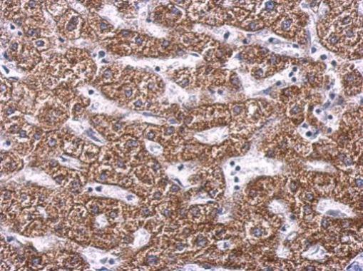
GTX101689 IHC-P Image
SDHA antibody detects SDHA protein at cytosol on human hepatoma by immunohistochemical analysis.
Sample: Paraffin-embedded hepatoma.
SDHA antibody (GTX101689) dilution: 1:500.
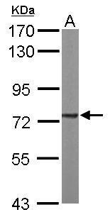
GTX101689 WB Image
Sample (50 ug of whole cell lysate)
A: mouse brain
7.5% SDS PAGE
GTX101689 diluted at 1:1000
The HRP-conjugated anti-rabbit IgG antibody (GTX213110-01) was used to detect the primary antibody.
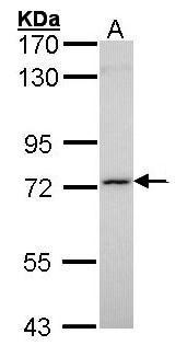
GTX101689 WB Image
Sample (30 ug of whole cell lysate)
A: Molt-4 (GTX27912)
7.5% SDS PAGE
GTX101689 diluted at 1:1000
The HRP-conjugated anti-rabbit IgG antibody (GTX213110-01) was used to detect the primary antibody.
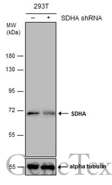
GTX101689 WB Image
Non-transfected (?) and transfected (+) 293T whole cell extracts (30 ug) were separated by 7.5% SDS-PAGE, and the membrane was blotted with SDHA antibody (GTX101689) diluted at 1:500. The HRP-conjugated anti-rabbit IgG antibody (GTX213110-01) was used to detect the primary antibody.