GTX101279 IHC-P Image
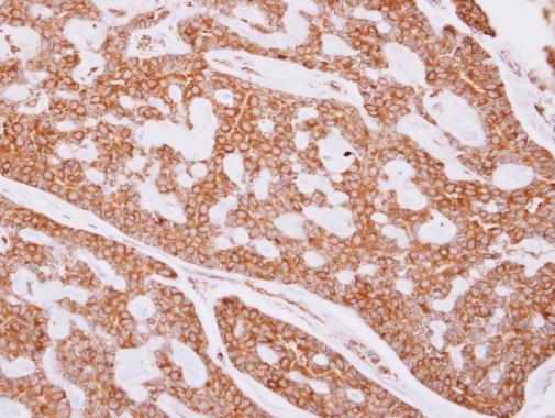
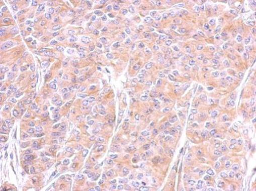
beta Tubulin antibody detects beta Tubulin protein at cytoplasm on human breast cancer by immunohistochemical analysis.
Sample: Paraffin-embedded breast cancer.
beta Tubulin antibody (GTX101279) dilution: 1:500.
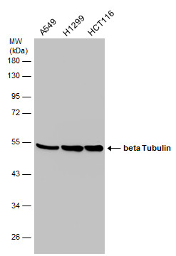
GTX101279 WB Image
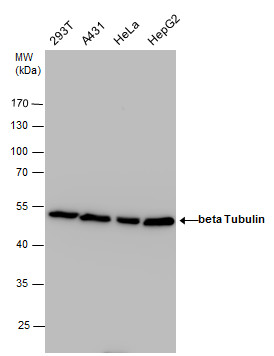
beta Tubulin antibody detects beta Tubulin protein by western blot analysis. Various whole cell extracts (30 ug) were separated by 10% SDS-PAGE, and the membrane was blotted with beta Tubulin antibody (GTX101279) diluted at a dilution of 1:3000. The HRP-conjugated anti-rabbit IgG antibody (GTX213110-01) was used to detect the primary antibody.
GTX101279 IHC-P Image
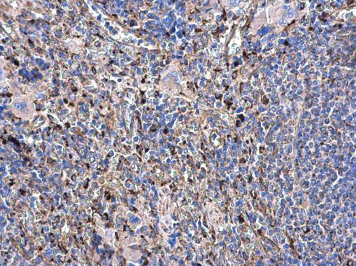
beta Tubulin antibody detects beta Tubulin protein at cytoplasm in rat spleen by immunohistochemical analysis.
Sample: Paraffin-embedded rat spleen.
beta Tubulin antibody (GTX101279) diluted at 1:500.
GTX101279 IHC-P Image
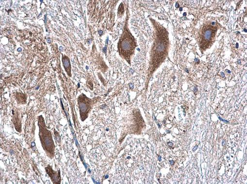
beta Tubulin antibody detects beta Tubulin protein at cytoplasm in rat spinal cord by immunohistochemical analysis.
Sample: Paraffin-embedded rat spinal cord.
beta Tubulin antibody (GTX101279) diluted at 1:500.
GTX101279 WB Image
Various whole cell extracts (30 ug) were separated by 10% SDS-PAGE, and the membrane was blotted with beta Tubulin antibody (GTX101279) diluted at 1:3000. The HRP-conjugated anti-rabbit IgG antibody (GTX213110-01) was used to detect the primary antibody.
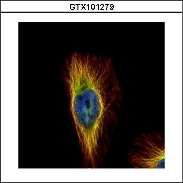
GTX101279 ICC/IF Image
Confocal immunofluorescence analysis (Olympus FV10i) of paraformaldehyde-fixed HeLa, using beta Tubulin(GTX101279) antibody (Green) at 1:500 dilution. Alpha-tubulin filaments were labeled with GTX11304 (Red) at 1:2000.
GTX101279 IHC-P Image
Immunohistochemical analysis of paraffin-embedded AGS xenograft, using beta Tubulin(GTX101279) antibody at 1:500 dilution.
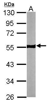
GTX101279 WB Image
Sample (30 ug of whole cell lysate)
A: NIH-3T3
10% SDS PAGE
GTX101279 diluted at 1:5000
The HRP-conjugated anti-rabbit IgG antibody (GTX213110-01) was used to detect the primary antibody.
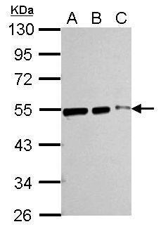
GTX101279 WB Image
Sample (whole cell lysate)
A: 293T 20ug
B: 293T 10ug
C: 293T 5ug
10% SDS PAGE
GTX101279 diluted at 1:10000
The HRP-conjugated anti-rabbit IgG antibody (GTX213110-01) was used to detect the primary antibody.