GTX101277 IHC-P Image
HMGB1 antibody detects HMGB1 protein at nucleus on mouse colon by immunohistochemical analysis.
Sample: Paraffin-embedded mouse colon.
HMGB1 antibody (GTX101277) dilution: 1:1000.
GTX101277 IHC-P Image
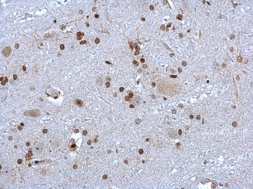
HMGB1 antibody detects HMGB1 protein at nucleus on rat brain stem by immunohistochemical analysis.
Sample: Paraffin-embedded rat brain stem.
HMGB1 antibody (GTX101277) dilution: 1:1000.
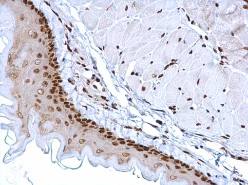
GTX101277 IHC-P Image
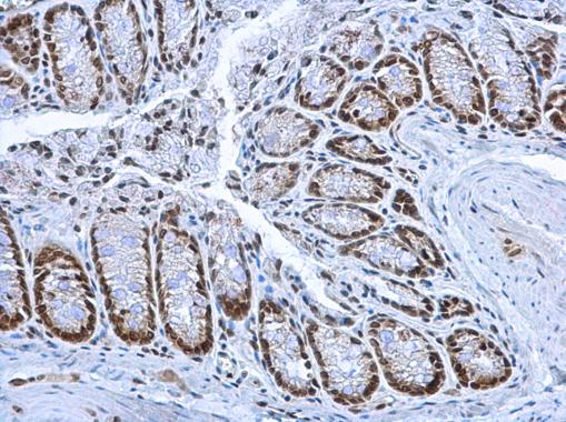
HMGB1 antibody detects HMGB1 protein at nucleus in mouse esophagus by immunohistochemical analysis.
Sample: Paraffin-embedded mouse esophagus.
HMGB1 antibody (GTX101277) diluted at 1:1000.
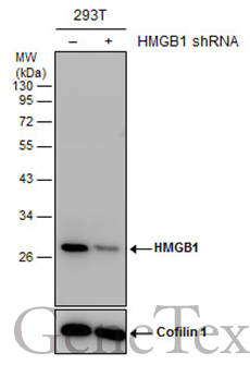
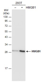
GTX101277 WB Image
Non-transfected (?) and transfected (+) 293T whole cell extracts (30 ug) were separated by 12% SDS-PAGE, and the membrane was blotted with HMGB1 antibody (GTX101277) diluted at 1:5000. The HRP-conjugated anti-rabbit IgG antibody (GTX213110-01) was used to detect the primary antibody.
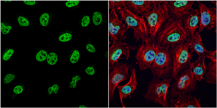
GTX101277 ICC/IF Image
HMGB1 antibody detects HMGB1 protein at nucleus by immunofluorescent analysis.
Sample: HeLa cells were fixed in 4% paraformaldehyde at RT for 15 min.
Green: HMGB1 protein stained by HMGB1 antibody (GTX101277) diluted at 1:1000.
Red: phalloidin, a cytoskeleton marker, stained by phalloidin (invitrogen, A12380) diluted at 1:200.
Blue: Hoechst 33342 staining.
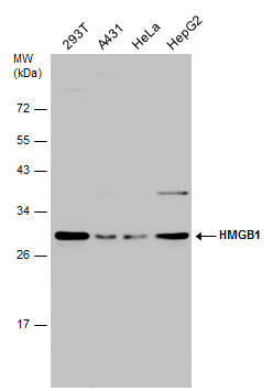
GTX101277 WB Image
Various whole cell extracts (30 ug) were separated by 12% SDS-PAGE, and the membrane was blotted with HMGB1 antibody (GTX101277) diluted at 1:3000.
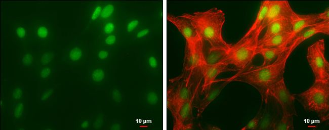
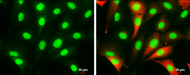
GTX101277 ICC/IF Image
HMGB1 antibody detects HMGB1 protein at nucleus by immunofluorescent analysis.
Sample: NIH/3T3 cells were fixed in 4% paraformaldehyde at RT for 15 min.
Green: HMGB1 protein stained by HMGB1 antibody (GTX101277) diluted at 1:500.
Red: phalloidin, a cytoskeleton marker, diluted at 1:50.
Scale bar = 10 um.
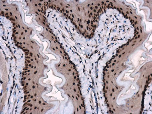
GTX101277 IHC-P Image
HMGB1 antibody detects HMGB1 protein at nucleus on mouse esophagus by immunohistochemical analysis.
Sample: Paraffin-embedded mouse esophagus.
HMGB1 antibody (GTX101277) dilution: 1:1000.
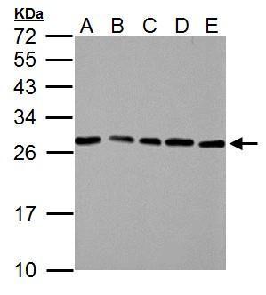
GTX101277 WB Image
HMGB1 antibody detects HMGB1 protein by western blot analysis.
A. 30 ug NIH-3T3 whole cell lysate/extract
B. 30 ug JC whole cell lysate/extract
C. 30 ug BCL-1 whole cell lysate/extract
D. 30 ug C2C12 whole cell lysate/extract
E. 30 ug Raw264.7 whole cell lysate/extract
12% SDS-PAGE
HMGB1 antibody (GTX101277) dilution: 1:3000
The HRP-conjugated anti-rabbit IgG antibody (GTX213110-01) was used to detect the primary antibody.
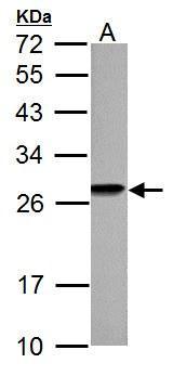
GTX101277 WB Image
HMGB1 antibody detects HMGB1 protein by western blot analysis.
A. 30 ug PC-12 whole cell lysate/extract
12% SDS-PAGE
HMGB1 antibody (GTX101277) dilution: 1:3000
The HRP-conjugated anti-rabbit IgG antibody (GTX213110-01) was used to detect the primary antibody.
GTX101277 ICC/IF Image
HMGB1 antibody detects HMGB1 protein at cytoplasm and nucleus by immunofluorescent analysis.
Sample: SK-N-SH cells were fixed in 4% paraformaldehyde at RT for 15 min.
Green: HMGB1 protein stained by HMGB1 antibody (GTX101277) diluted at 1:1000.
Red: beta Tubulin 3/ Tuj1, a cytoskeleton marker, stained by beta Tubulin 3/ Tuj1 antibody [GT11710] (GTX631836) diluted at 1:500.
Scale bar = 10 um.
GTX101277 WB Image
Non-transfected (?) and transfected (+) 293T whole cell extracts (30 ug) were separated by 12% SDS-PAGE, and the membrane was blotted with HMGB1 antibody (GTX101277) diluted at 1:5000. The HRP-conjugated anti-rabbit IgG antibody (GTX213110-01) was used to detect the primary antibody.