
GTX101127 IP Image
Lamin A + C antibody immunoprecipitates Lamin A + C protein in IP experiments.
IP samples: HeLa whole cell extract
A. 50 ug HeLa whole cell extract
B. Control with 4 ug of preimmune Rabbit IgG
C. Immunoprecipitation of Lamin A + C protein by 4 ug Lamin A + C antibody (GTX101127)
7.5 % SDS-PAGE
The immunoprecipitated Lamin A + C protein was detected by Lamin A + C antibody (GTX101127) diluted at 1:500.
[EasyBlot anti-rabbit IgG (GTX221666-01) was used as a secondary reagent]
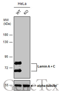
GTX101127 WB Image
Wild-type (WT) and Lamin A + C knockout (KO) HeLa cell extracts (30 ug) were separated by 7.5% SDS-PAGE, and the membrane was blotted with Lamin A + C antibody (GTX101127) diluted at 1:500. The HRP-conjugated anti-rabbit IgG antibody (GTX213110-01) was used to detect the primary antibody.
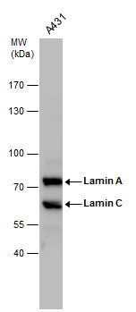
GTX101127 WB Image
Lamin A + C antibody detects Lamin A + C protein by western blot analysis. Whole cell extracts (30 ug) was separated by 7.5% SDS-PAGE, and the membrane was blotted with Lamin A + C antibody (GTX101127) diluted by 1:2000. The HRP-conjugated anti-rabbit IgG antibody (GTX213110-01) was used to detect the primary antibody.
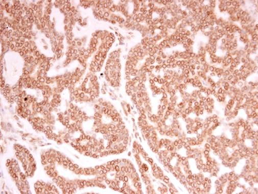
GTX101127 IHC-P Image
Lamin A + C antibody detects Lamin A + C protein at nuclear envelope in human breast carcinoma by immunohistochemical analysis.
Sample: Paraffin-embedded human breast carcinoma.
Lamin A + C antibody (GTX101127) diluted at 1:250.
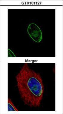
GTX101127 ICC/IF Image
Confocal immunofluorescence analysis (Olympus FV10i) of methanol-fixed HeLa, using Lamin A + C(GTX101127) antibody (Green) at 1:500 dilution. Alpha-tubulin filaments were labeled with GTX11304 (Red) at 1:500.

GTX101127 IHC-P Image
Immunohistochemical analysis of paraffin-embedded Mahlarvu xenograft, using Lamin A + C(GTX101127) antibody at 1:500 dilution.

GTX101127 IHC-P Image
Immunohistochemical analysis of paraffin-embedded C2C12 xenograft, using Lamin A + C(GTX101127) antibody at 1:500 dilution.

GTX101127 IHC-P Image
Immunohistochemical analysis of paraffin-embedded RT2 xenograft, using Lamin A + C(GTX101127) antibody at 1:500 dilution.

GTX101127 WB Image
Sample (30 ug of whole cell lysate)
A: NIH-3T3
7.5% SDS PAGE
GTX101127 diluted at 1:1000
The HRP-conjugated anti-rabbit IgG antibody (GTX213110-01) was used to detect the primary antibody.