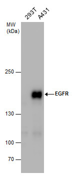
GTX100725 WB Image
EGFR antibody detects EGFR protein by western blot analysis. Whole cell extracts (30 ug) was separated by 5% SDS-PAGE, and the membrane was blotted with EGFR antibody (GTX100725) at a dilution of 1:1000. The HRP-conjugated anti-rabbit IgG antibody (GTX213110-01) was used to detect the primary antibody.
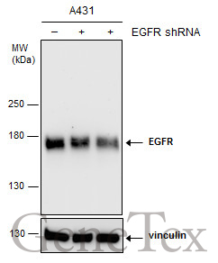
GTX100725 WB Image
Non-transfected (?) and transfected (+) A431 whole cell extracts (15 ug) were separated by 5% SDS-PAGE, and the membrane was blotted with EGFR antibody [N1], N-term (GTX100725) diluted at 1:3000. The HRP-conjugated anti-rabbit IgG antibody (GTX213110-01) was used to detect the primary antibody.
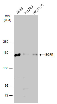
GTX100725 WB Image
Various whole cell extracts (30 ug) were separated by 7.5% SDS-PAGE, and the membrane was blotted with EGFR antibody [N1], N-term (GTX100725) diluted at 1:500. The HRP-conjugated anti-rabbit IgG antibody (GTX213110-01) was used to detect the primary antibody.
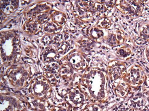
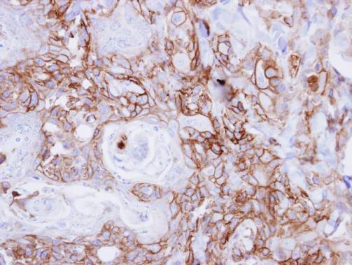
GTX100725 IHC-P Image
Immunohistochemical analysis of paraffin-embedded human breast cancer, using EGFR(GTX100725) antibody at 1:100 dilution.
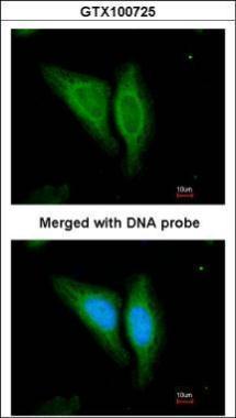
GTX100725 ICC/IF Image
Immunofluorescence analysis of paraformaldehyde-fixed HeLa, using EGFR(GTX100725) antibody at 1:200 dilution.
GTX100725 IHC-P Image
Immunohistochemical analysis of paraffin-embedded CA922 xenograft, using EGFR(GTX100725) antibody at 1:500 dilution.