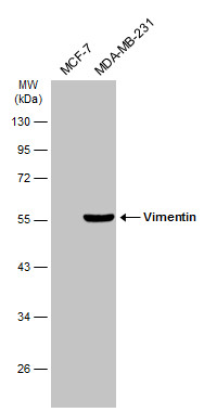
GTX100619 WB Image
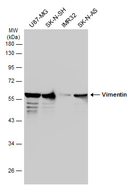
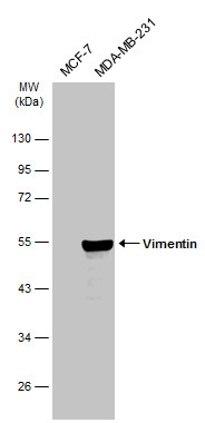
Various whole cell extracts (30 ug) were separated by 10% SDS-PAGE, and the membrane was blotted with Vimentin antibody (GTX100619) diluted at 1:50000. The HRP-conjugated anti-rabbit IgG antibody (GTX213110-01) was used to detect the primary antibody.
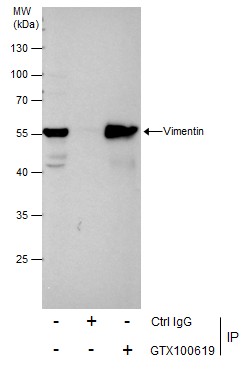
GTX100619 IP Image
Immunoprecipitation of Vimentin protein from HeLa whole cell extracts using 5 ug of Vimentin antibody (GTX100619).
Western blot analysis was performed using Vimentin antibody (GTX100619).
EasyBlot anti-Rabbit IgG (GTX221666-01) was used as a secondary reagent.
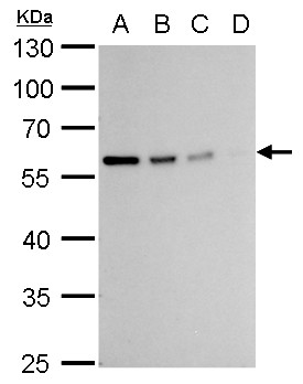
GTX100619 WB Image
Vimentin antibody detects Vimentin protein by western blot analysis.
A. 20 ug 293T whole cell lysate/extract
B. 10 ug 293T whole cell lysate/extract
C. 5 ug 293T whole cell lysate/extract
D. 1 ug 293T whole cell lysate/extract
10% SDS-PAGE
Vimentin antibody (GTX100619) dilution: 1:10000
The HRP-conjugated anti-rabbit IgG antibody (GTX213110-01) was used to detect the primary antibody.
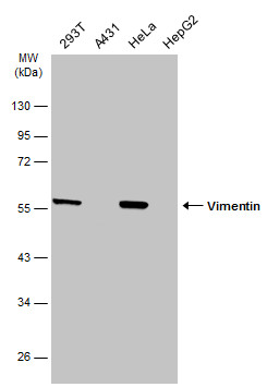
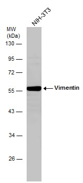
GTX100619 WB Image
Various whole cell extracts (30 ug) were separated by 10% SDS-PAGE, and the membrane was blotted with Vimentin antibody (GTX100619) diluted at 1:50000. The HRP-conjugated anti-rabbit IgG antibody (GTX213110-01) was used to detect the primary antibody.
GTX100619 WB Image
Whole cell extract (30 ug) was separated by 10% SDS-PAGE, and the membrane was blotted with Vimentin antibody (GTX100619) diluted at 1:2000.
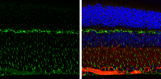
GTX100619 IHC-P Image
Vimentin antibody detects Vimentin protein expression by immunohistochemical analysis.
Sample:Paraffin-Embedded adult mouse retina.
Green: Vimentin protein stained by Vimentin antibody (GTX100619) diluted at 1:250.
Red: beta Tubulin 3/ TUJ1, stained by beta Tubulin 3/ TUJ1 antibody [GT11710] (GTX631836) diluted at 1:500.
Blue: Fluoroshield with DAPI (GTX30920).
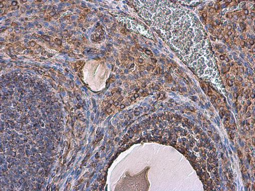
GTX100619 IHC-P Image
Vimentin antibody detects Vimentin protein at cell membrane and cytoplasm in rat ovary by immunohistochemical analysis.
Sample: Paraffin-embedded rat ovary.
Vimentin antibody (GTX100619) diluted at 1:500.
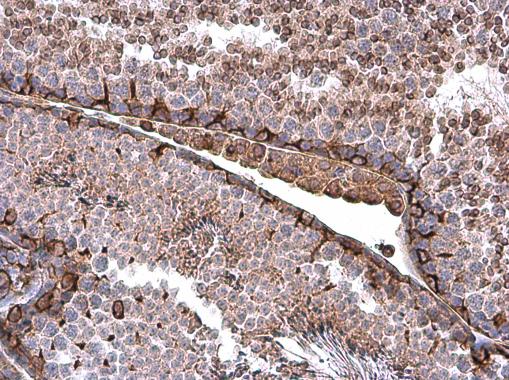
GTX100619 IHC-P Image
Vimentin antibody detects Vimentin protein at cell membrane and cytoplasm in mouse testis by immunohistochemical analysis.
Sample: Paraffin-embedded mouse testis.
Vimentin antibody (GTX100619) diluted at 1:500.
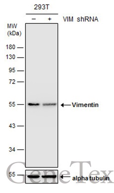
GTX100619 WB Image
Non-transfected (?) and transfected (+) 293T whole cell extracts (30 ug) were separated by 10% SDS-PAGE, and the membrane was blotted with Vimentin antibody (GTX100619) diluted at 1:20000.
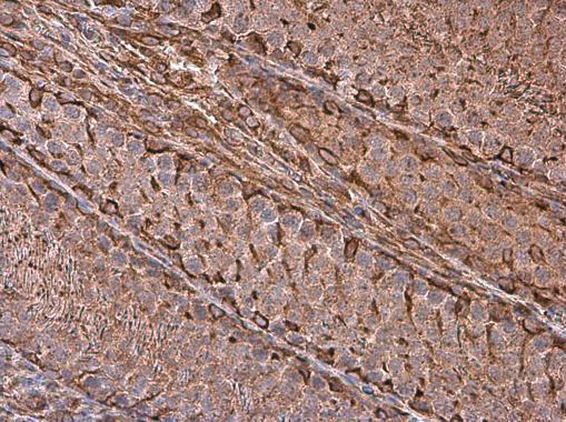
GTX100619 IHC-P Image
Vimentin antibody detects Vimentin protein at cell membrane and cytoplasm in rat testis by immunohistochemical analysis.
Sample: Paraffin-embedded rat testis.
Vimentin antibody (GTX100619) diluted at 1:500.
GTX100619 WB Image
Various whole cell extracts (30 ug) were separated by 10% SDS-PAGE, and the membrane was blotted with Vimentin antibody (GTX100619) diluted at 1:50000. The HRP-conjugated anti-rabbit IgG antibody (GTX213110-01) was used to detect the primary antibody.
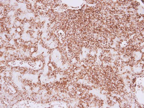
GTX100619 IHC-P Image
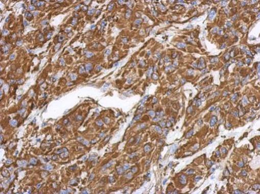
Vimentin antibody detects Vimentin protein at cytoplasm in human lung adenocarcinoma by immunohistochemical analysis.
Sample: Paraffin-embedded human lung adenocarcinoma.
Vimentin antibody (GTX100619) diluted at 1:500.
GTX100619 IHC-P Image
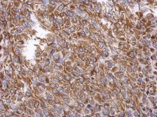
Immunohistochemical analysis of paraffin-embedded U373 xenograft, using Vimentin(GTX100619) antibody at 1:500 dilution.
GTX100619 IHC-P Image
Immunohistochemical analysis of paraffin-embedded RT2 xenograft, using Vimentin(GTX100619) antibody at 1:500 dilution.
GTX100619 ICC/IF Image
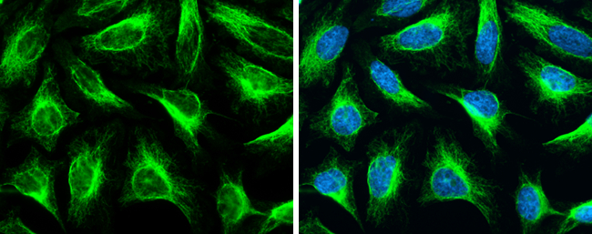
Vimentin antibody detects Vimentin protein at cytoskeleton by immunofluorescent analysis.Sample: HeLa cells were fixed in 4% paraformaldehyde at RT for 15 min.Green: Vimentin stained by Vimentin antibody (GTX100619) diluted at 1:500.Blue: Hoechst 33342 staining.
GTX100619 WB Image
Various whole cell extracts (30 ug) were separated by 10% SDS-PAGE, and the membrane was blotted with Vimentin antibody (GTX100619) diluted at 1:10000. The HRP-conjugated anti-rabbit IgG antibody (GTX213110-01) was used to detect the primary antibody.