GTX100496 IHC-P Image
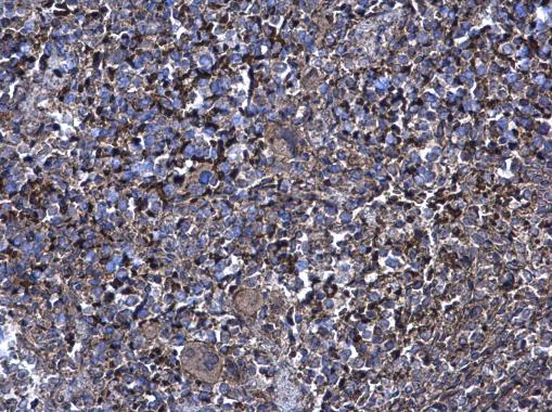
XIAP antibody [C1C3] detects XIAP protein at cytoplasm on mouse spleen by immunohistochemical analysis.
Sample: Paraffin-embedded mouse spleen.
XIAP antibody [C1C3] (GTX100496) diluted at 1:500.
GTX100496 IHC-P Image
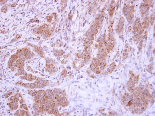
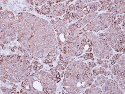
XIAP antibody [C1C3] detects XIAP protein at cytoplasm in human breast carcinoma by immunohistochemical analysis.
Sample: Paraffin-embedded human breast carcinoma.
XIAP antibody [C1C3] (GTX100496) diluted at 1:500.
GTX100496 WB Image
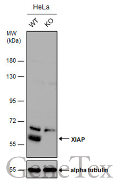
Wild-type (WT) and XIAP knockout (KO) HeLa cell extracts (30 ug) were separated by 7.5% SDS-PAGE, and the membrane was blotted with XIAP antibody [C1C3] (GTX100496) diluted at 1:500. The HRP-conjugated anti-rabbit IgG antibody (GTX213110-01) was used to detect the primary antibody.
GTX100496 WB Image
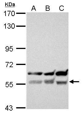
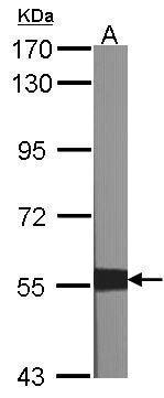
XIAP antibody [C1C3] detects XIAP protein by western blot analysis.
A. 30 ug 293T whole cell lysate/extract
B. 30 ug A431 whole cell lysate/extract
C. 30 ug H1299 whole cell lysate/extract
7.5% SDS-PAGE
XIAP antibody [C1C3] (GTX100496) dilution: 1:500
The HRP-conjugated anti-rabbit IgG antibody (GTX213110-01) was used to detect the primary antibody.
GTX100496 IHC-P Image
Immunohistochemical analysis of paraffin-embedded SW480 xenograft, using BIRC4(GTX100496) antibody at 1:500 dilution.
GTX100496 WB Image
Sample (30 ug of whole cell lysate)
A:NIH-3T3
7.5% SDS PAGE
GTX100496 diluted at 1:1000
The HRP-conjugated anti-rabbit IgG antibody (GTX213110-01) was used to detect the primary antibody.
GTX100496 ICC/IF Image
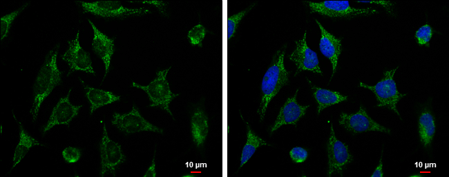
XIAP antibody [C1C3] detects XIAP protein at cytoplasm by immunofluorescent analysis.
Sample: HeLa cells were fixed in ice-cold MeOH for 5 min.
Green: XIAP protein stained by XIAP antibody [C1C3] (GTX100496) diluted at 1:500.
Blue: Hoechst 33342 staining.
Scale bar = 10 um.